

Физиология растений, 2023, T. 70, № 7, стр. 858-865
Накопление полифенолов и нафтохинонов в морфогенных культурах двух видов рода Drosera
А. В. Моршнева a, b, *, М. Т. Ханды a, b, В. П. Григорчук a, Г. К. Чернодед a, Т. Ю. Горпенченко a
a Федеральный научный центр биоразнообразия наземной биоты Восточной Азии ДВО РАН
Владивосток, Россия
b Дальневосточный федеральный университет
Владивосток, Россия
* E-mail: morshneva.av@students.dvfu.ru
Поступила в редакцию 05.09.2023
После доработки 07.11.2023
Принята к публикации 11.11.2023
- EDN: ZFGIDI
- DOI: 10.31857/S0015330323600717
Аннотация
Получена ризогенная культура Drosera capensis L. Методом ВЭЖХ-УФ-МС проведен сравнительный анализ влияния разных методов сушки на показатель выхода индивидуальных полифенолов и 1,4-нафтохинонов ризогенной культуры D. capensis L. с ранее полученной морфогенной длительно-растущей (более 15 лет) культурой D. rotundifolia L. В ризогенной культуре D. capensis L. впервые идентифицировано 6 соединений (мирицетин-3-O-β-глюкопиранозид, россолизид, 3,3'-ди-O-метилэллаговой кислоты 4-О-β-D-гликопиранозид, мирицетин, 3,3'-ди-O-метилэллаговая кислота, плюмбагин).
ВВЕДЕНИЕ
Росянка (Drosera L.) является одним из крупнейших родов плотоядных растений из семейства росянковых (Droseraceae). Эти растения широко применяются в народной медицине, а их экстракты обладают антимикробными, противогрибковыми и противоопухолевыми свойствами [1, 2]. Считается, что биологическая активность Drosera обусловлена 1,4-нафтохинонами и флавоноидами [3].
Разные виды Drosera накапливают два основных нафтохинона – плюмбагин и 7-метилюглон [4], и в небольших количествах – гидроксиплюмбагин-4-О-гликозида, дрозерон, гидроксидрозерон, дрозерон-5-гликозид, гидроксидрозерон-8-гликозид, россолизид [3, 5–7]. Основными флавоноидами, обнаруженными в роде Drosera, являются кверцетин, гиперозид, кемпферол, мирицетин [8–10]. Еще одной группой вторичных метаболитов Drosera являются эллаготанины, в частности, продукт их разложения – эллаговая кислота [11]. Таким образом, род Drosera – богатый источник экономически ценных вторичных метаболитов. Однако, естественный ареал росянки под действием антропогенных факторов стремительно сокращается [12]. Получение хорошо растущей культуры клеток Drosera с высокими показателями синтеза биологически активных веществ востребовано на рынке фармакологических субстанций [13].
Исследование вторичного метаболизма в целом растении весьма трудоемкий процесс, обусловленный долгим жизненным циклом растений, привязанностью к месту обитания, климату, зависимостью от погодных и других условий окружающей среды. Одним из эффективных способов в исследовании биохимических процессов и самих веществ является использование модельных систем. Для растений, одним из вариантов модельных систем является культура клеток, тканей и органов in vitro. Поэтому культура клеток росянки является не только источником ценных вторичных метаболитов, но и важным объектом в исследовании и в понимании вторичного метаболизма полифенолов и нафтохинонов. Различные природные модификации структур молекул определяют специфические свойства веществ. Способ извлечения этих веществ, применяемый растворитель и метод сушки растительной ткани в итоге могут негативно сказываться на составе вторичных метаболитов растительных тканей.
Цель данной работы – исследовать состав полифенолов и нафтохинонов в морфогенных культурах двух видов D. rotundifolia и D. capensis при различном способе высушивания исходного материала.
МАТЕРИАЛЫ И МЕТОДЫ
Объект исследования. В качестве объектов исследования использовали две линии морфогенной культуры D. rotundifolia под обозначением “DR Green Dark”, “DR Green Light”, полученные в 2005 г. в лаборатории биотехнологии Биолого-почвенного института [14], и полученную в рамках настоящего исследования ризогенную культуру in vitro D. capensis под обозначением “DC Dark”.
Получение культур клеток D. capensis. В качестве эксплантов использовали листья растения Drosera capensis L. Листья промывали детергентом, затем теплой водой и стерилизовали 0.1% диоцидом в течение 5 мин. После стерилизации материал трехкратно отмывали в стерильной дистиллированной воде. Стерильные листья просушивали на стерильной фильтровальной бумаге и использовали в качестве эксплантов, помещая их на агаризованную питательную среду. Чашки Петри с первичным эксплантом культивировали в темноте при температуре 25 ± 1°С и влажности 70 ± 5%.
Для индукции каллусогенеза использовали MС-среду, приготовленную по стандартной прописи [15] c семью разными сочетаниями фитогормонов: α-нафтилуксусную кислоту (НУК), кинетин (кин) и 6-бензиламинопурин (БАП). Сочетания и концентрации гормонов в среде представлены в табл. 1.
Таблица 1.
Гормональный состав сред, использованных при получении и выращивании культуры клеток D. capensis
| № среды | 1 | 2 | 3 | 4 | 5 | 6 | 7 |
|---|---|---|---|---|---|---|---|
| Гормон и его концентрация в среде, мг/л | НУК 2/кин 1 | НУК 2/кин 0.5 | НУК 1/кин 0.5 | НУК 2 | НУК 2/БАП 1 | НУК 2/БАП 0.5 | НУК 1/БАП 0.5 |
Инициация образования каллуса произошла при замене регулятора роста на 2,4-дихлорфеноксиуксусную кислоту (2,4-Д) в концентрации 1 мг/л.
После начала каллусогенеза объекты были перенесены на стандартную коллекционную среду W0 (MС-среда со сниженным содержанием нитрата аммония NH4NO3 до 400 мг/л среды), используемую в институте (ФНЦ Биоразнообразия ДВО РАН), с регуляторами роста НУК (2 мг/л) и БАП (0.5 мг/л).
Исследование ростовых характеристик полученной линии D. capensis и двух других исследуемых линий D. rotundifolia производили на основании данных сухого веса биомассы. Индекс роста определяли по формуле:
где ${{W}_{0}}$ ‒ начальная масса культуры, г; ${{W}_{t}}$ ‒ масса культуры в конце цикла выращивания, г.Условия культивирования. Культивирование проводили на среде W0 с добавлением тиамина (B1) ‒ 0.2 мг/л, пиридоксина (B6) ‒ 0.5 мг/л, никотиновой кислоты (PP) ‒ 0.5 мг/л, мезоинозитола ‒ 100 мг/л, пептона ‒ 100 мг/л, сахарозы ‒ 25000 мг/л, агара ‒ 6000 мг/л и гормонов НУК ‒ 2.0 мг/л, БАП ‒ 0.5 мг/л. Каллусы выращивали в колбах объемом 100 мл (33 мл питательной среды) в темноте (линии D. capensis “DC Dark”, D. rotundifolia “DR Green Dark”) при 25 ± 1°С и на свету (линия D. rotundifolia “DR Green Light”) при 25 ± 1°С, влажности 70 ± 5% и интенсивности освещения люминесцентными лампами (белый свет) 49 мкмоль/(с м2) в режиме 16/8 ч день/ночь, соответственно. Цикл выращивания составлял 30 сут. Для пересева использовали фрагменты каллуса с размером 0.8 см3.
Подготовка проб для фитохимического анализа. Для анализа биомассу подготовили двумя разными способами: высушивали в потоке теплого воздуха (+40°С) и измельчили с использованием жидкого азота (1 способ), высушивали в аппарате лиофильной сушки (2 способ). 50 мг измельченной биомассы экстрагировали 1 мл 80% метанола в течение 30 мин с использованием ультразвуковой бани. Экстракт выдерживали 12 ч без доступа света, супернатант фильтровали (0.45 мкм, Millipore, “Бедфорд”, США) и использовали для фитохимического анализа.
Фитохимический анализ. Для анализа вторичных метаболитов в биомассе морфогенных культур D. rotundifolia и D. capensis была использована аналитическая высокоэффективная жидкостная хроматография с ультрафиолетовым и масс-спектрометрическим детектированием (ВЭЖХ-УФ-МС). Экстракты анализировали с использованием хроматографа 1260 Infinity (“Agilent”, США) совмещенного с тандемным масс-спектрометром Bruker HCT ultra PTM Discovery System (“Bruker Daltonik”, GmbH, Германия). Разделение проводили на аналитической колонке Zorbax C18 (150 × 2.1 мм, 3.5 мкм, “Agilent”, США) термостатированной при 40°С. В качестве подвижной фазы использовали раствор муравьиной кислоты (0.1%) в деионизированной воде (раствор А) и ацетонитрил (раствор Б). Градиентное элюирование проводили со скоростью потока 0.2 мл/мин по следующей схеме: от 10% до 35% раствора Б за 40 мин, затем до 95% раствора Б до 50 мин и 95% раствора Б до 60 мин. УФ-спектры записывали в диапазоне λ 200–800 нм с использованием детектора на диодной матрице. Масс-спектрометрические данные для подтверждения идентификации определяемых компонентов получали в условиях ионизации электрораспыления и регистрации отрицательных ионов с диапазоном регистрируемых значений m/z 100–1000. Количественный анализ полифенолов производили при длине волны 265 нм, нафтохинонов – при 330 нм.
Статистический анализ данных. Для статистической обработки данных использовали программу GraphPad Prism 8. Достоверность результатов была рассчитана с помощью t-теста, критерия Уилкоксона и U-критерия Манна-Уитни.
РЕЗУЛЬТАТЫ
Получение культур клеток D. capensis. На первых этапах работы с D. capensis, основываясь на литературных данных, для индукции каллусогенеза использовали агаризованную MС-среду с НУК и цитокининами [16, 17]. Культивирование эксплантов на этих средах в течение двух месяцев существенных результатов не дало. При повторных экспериментах, для индукции каллусогенеза регулятор роста поменяли на 2,4-Д. В результате проведенных экспериментов по влиянию различных регуляторов роста на каллусогенез было установлено, что первичная культура D. capensis формируется на среде, содержащей гормон 2,4-Д в концентрации 1 мг/мл. Первичный каллусогенез начинался непосредственно из листьев спустя пятнадцать дней культивирования с образованием побегов. Полученные каллусные культуры характеризовались светло-желтым цветом и твердой структурой. После переноса культур на среду W0 НУК 2 мг/л, БАП 0.5 мг/л, на двадцатые сутки был сформирован твердый ризогенный каллус in vitro с плотной структурой (рис. 1в). На некоторых участках образовывались плотные перьевидные отростки белого цвета. На остальных вариантах используемых сред ростовые процессы не были выявлены. Полученная культура выращивается в течение 2 лет, за это время ее морфологическая структура не менялась. Индекс роста полученной культуры D. capensis составляет 3.7. Индекс роста линий D. rotundifolia “DR Green Dark” “DR Green Light” равен 2.2 и 5.0, соответственно.
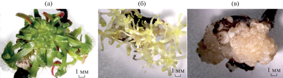
Рис. 1.
Внешний вид линий морфогенных культур D. rotundifolia и D. capensis: (а) – линия “DR Green Light”; (б) – “DR Green Dark”; (в) – “DC Dark”. Масштаб 1:5.
Фитохимическая характеристика морфогенных культур Drosera. В результате ВЭЖХ-УФ-МС анализа в исследуемых линиях идентифицировано двенадцать биологически-активных соединений – флавоноиды и их гликозиды (мирицетин-3-O-β-глюкопиранозид (два изомера), гиперозид, изокверцетин, мирицетин и кверцетин), производные эллаговой кислоты (гликопиранозид 3-O-метилэллаговой кислоты, 3,3'-ди-О-метилэллаговой кислоты 4-O-β-D-гликопиранозид, 3,3'-ди-O-метилэллаговой кислоты), галлат эпигаллокатехина и 1,4-нафтохиноны (россолизид и плюмбагин) (рис. 2). Идентификация соединений была произведена ранее и опубликована в нашей предыдущей работе [14].
Рис. 2.
Хроматографический профиль (ВЭЖХ, 330 нм) экстрактов из морфогенных каллусов D. rotundifolia и D. capensis после сушки теплым воздухом (+40°С): (а) – линия “DR Green Light”; (б) – “DR Green Dark”; (в) – “DC Dark”. Нумерация на хроматограмме соответствует номерам пиков из таблицы 2.

В ризогенном каллусе in vitro D. capensis идентифицировано 6 основных соединений: мирицетин-3-O-β-глюкопиранозид, россолизид, 3,3'-ди-O-метилэллаговая кислота и ее 4-О-β-D-гликопиранозид, мирицетин, плюмбагин (рис. 2).
Сопоставление разных способов сушки и использования свежесобранной биомассы для количественного анализа полифенолов и 1,4-нафтохинонов выявило несколько различий (табл. 2). Первое, при разных методах сушки качественный состав полифенолов существенно не различался (за исключением галлата эпикатехина, который не был обнаружен при воздушной сушке в двух линиях), однако, количественное содержание при лиофильной сушке было выше (от 0.01 до 0.2 мкг/г сухого веса). Второе, 1,4-нафтохиноны лучше идентифицировались после воздушной сушки. Поскольку 1,4-нафтохиноны являются, наравне с флавоноидами, одними из важных целевых соединений Drosera определяющих биологическую активность экстрактов, для дальнейших расчетов и оценки продуктивности линий использовали результаты анализа экстрактов образцов после воздушной сушки.
Таблица 2.
Количественное содержание вторичных метаболитов в экстрактах морфогенных культур Drosera, высушенных на воздухе, с помощью лиофильной сушки и из свежей ткани
| Вид сушки | Воздушная сушка (+40°С) | Лиофильная сушка | |||||
|---|---|---|---|---|---|---|---|
| линия | “DR Green Light” | “DR Green Dark” | “DC Dark” | “DR Green Light” | “DR Green Dark” | “DC Dark” | |
| номер пика на хроматограмме | соединение | мкг/г сухого веса | мкг/г сухого веса | мкг/г сухого веса | мкг/г сухого веса | мкг/г сухого веса | мкг/г сухого веса |
| 1 | Галлат эпигаллокатехина | + | 0 | 0 | + | + | + |
| Флавоноиды | |||||||
| 2 | Мирицетин-3-O-β-глюкопиранозид I | 2.184 | 0.495 | 0.227 | 2.058 | 0.464 | 0.395 |
| 3 | Мирицетин-3-O-β-глкопиранозид II | 0.46 | 0 | 0 | 0.203 | 0 | 0 |
| 6 | Гиперозид | 6.65 9 | 1.547 | 0 | 6.739 | 1.757 | 0 |
| 7 | Изокверцетин | 1.182 | 0.225 | 0 | 1.391 | 0.457 | 0 |
| 9 | Мирицетин | 1.037 | 0.505 | 0.256 | 1.198 | 0.496 | 0 |
| 10 | Кверцетин | 3.589 | 1.694 | 0 | 4.065 | 1.729 | 0 |
| Производные эллаговой кислоты | |||||||
| 5 | Гликопиранозид 3-O- метилэллаговой кислоты | 0.567 | 0.156 | 0 | 0.771 | 0 | 0 |
| 8 | 3,3'-ди-О- метилэллаговой кислоты 4-O-β-D- гликопиранозид | 1.51 | 0.645 | 0.281 | 1.712 | 0.601 | 0.54 |
| 11 | 3,3’-ди- O-метилэллаговая кислота | 0.551 | 0.847 | 0.549 | 0.82 | 0.944 | 0.347 |
| Нафтохиноны | |||||||
| 4 | Россолизид | + | + | + | + | + | 0 |
| 12 | Плюмбагин | 0 | 0.06 | 0.111 | 0 | 0.125 | 0 |
Количественный анализ полифенолов и нафтохинонов, содержащихся в культурах клеток двух видов растения рода Drosera – D. rotundifolia и D. capensis выявил существенные различия между линиями (табл. 2). Высокие показатели производительности полифенолов установлены для линии “DR Green Light”, в частности, по накоплению флавоноидов, они превышают в три раза аналогичные показатели в линии “DR Green Dark”, и в 20 раз – линии “DC Dark”. По показателям производных эллаговой кислоты в 1.5 раза – линии “DR Green Dark” и в 3 раза “DC Dark”. По показателям накопления 1,4-нафтохинонов положительно отличилась полученная линия ризогенного каллуса D. capensis “DC Dark”. В этом случае, показатель плюмбагина в два раза превышает все линии “DR Green Dark”.
ОБСУЖДЕНИЕ
Получение новых культур клеток является неотъемлемым этапом большинства направлений исследований растительной биологии и биотехнологии. Особый интерес при этом представляют культуры клеток плотоядных растений, как источники ценных вторичных метаболитов. Получение микроклонов и культивирование растений Drosera хорошо разработанная технология [17]. Однако, плантационное культивирование D. rotundifolia и D. capensis практически невозможно. Во-первых, растения сами по себе маленького размера 10–15 см в высоту, во-вторых, специфичная среда обитания – торфяные болота, площадей которых с каждым годом все меньше [12], а воссоздание среды обитания – затратное мероприятие. Полученная ранее нами стабильная длительно-растущая морфогенная культура D. rotundifolia дала основу для поиска новых систем для исследования вторичного метаболизма плотоядных растений. D. capensis, как близкородственного вида D. rotundifolia со схожим фитохимическим составом, могла бы стать новой сравнительной моделью.
Получить каллусную культуру D. capensis удалось с использованием гормона 2,4-Д из листьев интактного растения классическими методами. Индукция каллусогенеза является известным свойством 2,4-Д [18, 19]. Длительное выращивание на 2,4-Д оказалось невозможным из-за ингибирования ростовых процессов на втором пассаже. В результате произвели смену среды на стандартную коллекционную среду с фитогормонами НУК и БАП, которая привела к получению ризогенной культуры с высокими показателями роста (индекс роста не менее 5 в течение года). Полученный результат интересен тем, что при микроклональном размножении растений рода Drosera весьма проблематичным является индукция корнеобразования и, именно, укоренение растения [17, 20–22], а в нашем случае сразу происходит формирование ризогенной клеточной культуры. Использование данного соотношения гормонов может быть перспективным и для укоренения микроклонированных растений. В аналогичных условиях ранее была получена морфогенная клеточная культура D. rotundifolia, культивируемая длительное время (с 2005 г.) в темноте, однако, ткань развивалась преимущественно побегами [14]. При выращивании морфогенной клеточной культуры D. rotundifolia на свету повышались показатели накопления вторичных метаболитов – флавоноидов и 1,4-нафтохинонов, при этом показатели роста были выше, чем у тканей, выращиваемых в темноте. Свет является одним из наиболее важных факторов, влияющих на рост и выработку вторичных метаболитов растениями [23]. В исследовании о влиянии светодиодных источников света на D. bermannii было показано, что при белом светодиодном освещении увеличивалось количество плюмбагина в побегах относительно корней, но спустя две недели эффект прошел [24]. Напротив, в работе Wojciech с соавт. [25] о влиянии сине-красного света на фенольные соединения в семействе Droseraceae полагают, что этот фактор не изменяет общего содержания фенолов, но влияет на накопление специфических фенолов и снижает содержание плюмбагина. На основании имеющихся данных можно заключить, что вопрос влияния света на жизнедеятельность росянки, в частности на накопление вторичных метаболитов, остается дискуссионным.
Исследование особенностей накопления и извлечения биологически активных вторичных метаболитов из культур клеток является важной прикладной и фундаментальной задачей современной биологии растений. В растениях in vitro Drosera, обнаружены флавоноиды: кверцетин, кверцетрин, изокверцетин, мирицетрин и др. в небольшом количестве в видах D. rotundifolia, D. capensis, а также D. intermedia, D. spatulata [21]. Кроме самих флавоноидов, в растениях находят их гликозилированные формы, наиболее распространенными из которых являются формы 3-О-глюкозида или 3-О-галактозида. Эти вещества распространены во многих видах растений. Например, у D. madagascariensis и D. rotundifolia было обнаружено, что кверцетин составляет около 2 и 0.49% сухого веса исследованных растений соответственно, тогда как количество флавонолгликозида – гиперозида (кверцетин 3-O-галактозид) составляло 3.75 и 6.36%, соответственно [26, 27]. Другое исследование D. rotundifolia также подтвердило эту тенденцию, выявив 0.17 и 2.89% сухого веса для кверцетина и гиперозида, соответственно [9]. В нашей работе наблюдается такая же закономерность (табл. 2). Количественное содержание флавоноидных гликозидов варьирует относительно литературных данных. Так, общее содержание веществ в среднем составляет от 5 до 10%, при этом среди флавоноидов обнаружены чаще всего: гиперозид, мирицетин-3-О-галактозид, изокверцитрин, кверцетин. В исследовании Paul A. Egan [3] показано, что в D. rotundifolia, кверцетин может быть обнаружен на уровне 6.40%, а кверцетин-3-О-галактозид – на уровнях, достигающих 6.36%. Большее количество флавоноидов и их гликозидов находится в листьях, стеблях и цветках растений, чем в корнях [17]. В нашей работе флавоноиды также преобладали в морфогенной культуре, состоящей из надземных побегов.
В литературных данных сообщается, что интактное растение D. rotundifolia богато флавоноидами, 1,4-нафтохинонами, эллаговой кислотой и ее производными [28]. Большинство из этих веществ были идентифицированы в исследуемых образцах. Их структурное разнообразие схоже описанному в литературе для интактных растений D. rotundifolia и количественно больше в образцах линии “DR Green Light” [28, 29]. В исследовании R. Caniato с соавт. [30] сообщается об отсутствии вариации соединений между корнями и надземными органами у диких популяций D. rotundifolia, у видов D. capensis наблюдается заметная разница между содержанием нафтохинонов в корнях и побегах растений – в корнях большее содержание 7-метилюглона, чем в побегах данного вида. Однако был проведен отдельный анализ подборки частей растений D. capensis, который показал, что самая большая концентрация 7-метилюглона была обнаружена в цветках.
Выделение и биосинтез галлат эпигаллокатехина мало рассмотрен и обобщен в литературных данных о хищных растениях. Однако, это соединение является основным компонентом различных видов чая, содержащимся преимущественно в листьях и проявляющем значительную биоактивность, включая потенциальную противораковую и противовоспалительную [31]. Малая изученность биосинтеза и физиологических функций этого соединения у хищных растений может открыть новое направление для исследований.
В нашем исследовании содержание флавоноидных гликозидов, производных эллаговой кислоты и 1,4-нафтохинонов значительно не отличается, однако измельчение высушенного теплым воздухом материала в ступке более трудоемко, чем высушенного лиофилизацией.
Таким образом, исследуемые морфогенные культуры D. rotundifolia и D. capensis являются перспективными источниками возобновляемого сырья с высоким содержанием полифенолов и 1,4-нафтохинонов. При этом способ высушивания материала лишь незначительно влияет на выход исследуемых веществ.
В работе использовали оборудование Центра коллективного пользования “Биотехнология и генетическая инженерия” ФНЦ Биоразнообразия ДВО РАН.
Работа выполнена в рамках государственного задания Министерства науки и высшего образования Российской Федерации (№ 121031000144-5) и при частичной поддержке Российской федеральной программы академического лидерства “Приоритет 2030” (№ 23-01-3.04.0001).
Настоящая статья не содержит каких-либо исследований с участием людей и животных в качестве объектов исследования. Авторы заявляют об отсутствии конфликта интересов.
Список литературы
Didry N., Dubreuil L., Trotin F., Pinkas M. Antimicrobial activity of aerial parts of Drosera peltata Smith on oral bacteria // J. Ethnopharm. 1998. V. 60. P. 91. https://doi.org/10.1016/S0378-8741(97)00129-3
Crowder A.A., Pearson M.C., Grubb P.J., Langlois P.H. Drosera L. // J. Ecol. 1990. V. 78. P. 233. https://doi.org/10.2307/2261048
Egan P.A., Kooy F. Phytochemistry of the carnivorous sundew genus Drosera (Droseraceae) – future perspectives and ethnopharmacological relevance // Chem. Biodiversity. 2013. V. 10. P. 1774. https://doi.org/10.1002/cbdv.201200359
Kämäräinen T., Uusitalo J., Jalonen J., Laine K., Hohtola A. Regional and habitat differences in 7-methyljuglone content of Finnish Drosera rotundifolia // Phytochem. 2003. V. 63. P. 309. https://doi.org/10.1016/S0031-9422(03)00115-8
Marczak L., Kawiak A., Lojkowska E., Stobieck M. Secondary metabolites in in vitro cultured plants of the genus Drosera // Phytochem. Anal. 2005. V. 16. P. 143. https://doi.org/10.1002/pca.833
Budzianowski J., Skrzypczak L., Kukulczanka K. Phenolic compounds of Drosera intermedia and D. spathulata from in vitro cultures // Acta Hortic. 1993. V. 330. P. 277. https://doi.org/10.17660/ActaHortic.1993.330.36
Gu J.Q., Graf T.N., Lee D., Chai H.B., Mi Q., Kardono L.B.S., Setyowati F.M., Ismail R., Riswan S., Farnsworth N.R., Cordell G.A., Pezzuto J.M., Swanson S.M., Kroll D.J., Falkinham J.O., et al. Cytotoxic and antimicrobial constituents of the bark of Diospyros maritima collected in two geographical locations in Indonesia // J. Nat. Prod. 2004. V. 67. P. 1156. https://doi.org/10.1021/np040027m
Zehl M., Braunberger C., Conrad J., Crnogorac M., Krasteva S., Vogler B., Beifuss U., Krenn L. Identification and quantification of flavonoids and ellagic acid derivatives in therapeutically important Drosera species by LC-DAD, LC-NMR, NMR, and LC-MS // Anal. Bioanal. Chem. 2011. V. 400. P. 2565. https://doi.org/10.1007/s00216-011-4690-3
Fukushimaa K., Nagai K., Hoshi Y., Masumoto S., Mikami I., Takahashi Y., Oike H., Kobori M. Drosera rotundifolia and Drosera tokaiensis suppress the activation of HMC-1 human mast cells // J. Ethnopharm. 2009. V. 125. P. 90. https://doi.org/10.1016/j.jep.2009.06.009
Sprague-Piercy M.A., Bierma J.C., Crosby M.G. The droserasin 1 PSI: a membrane-interacting antimicrobial peptide from the carnivorous plant Drosera capensis // Biomolec. 2020. V. 10. P. 1069. https://doi.org/10.3390/biom10071069
Sharifi-Rad J., Quispe C., Castillo C.M.S., Caroca R., Lazo-Vélez M.A., Antonyak H., Polishchuk A., Lysiuk R., Oliinyk P., Masi L.D., Bontempo P., Martorell M., Daştan S.D., Rigano D., Wink M. et al. Ellagic acid: a review on its natural sources, chemical stability, and therapeutic potential // Oxid. Med. Cell. Longevity. 2022. V. 2022. P. 24. https://doi.org/10.1155/2022/3848084
Barthlott W., Hostert A., Kier G., Küper W., Kreft H., Mutke J., Rafiqpoor M.D., Sommer J.H. Geographic patterns of vascular plant diversity at continental to global scales // Sitzungsber. Akad. Wiss. Wien, Math.-Naturwiss. Kl., Abt. 1. 2007. V. 61. P. 305. https://doi.org/10.3112/erdkunde.2007.04.01
Baranyai B., Joosten H. Biology, ecology, use, conservation and cultivation of round-leaved sundew (Drosera rotundifolia L.): a review // Mires Peat. 2016. V. 18. P. 1. https://doi.org/10.19189/MaP.2015.OMB.212
Khandy M.T., Chernoded G.K., Grigorchuk V.P., Vereshchagina Yu.V., Morshneva A.V., Gorpenchenko T.Yu. Histological structure and composition of secondary metabolites in cell culture of Drosera rotundifolia L. // Russ. J. Plant Physiol. 2022. V. 69. P. 451. https://doi.org/10.1134/S1021443722050090
Murashige T., Skoog F. A revised medium for rapid growth and bio assays with Tobacco tissue cultures // Physiol. Plant. 1962. V. 15. P. 473. https://doi.org/10.1111/j.1399-3054.1962.tb08052.x
Crouch I.J., Staden J. In vitro propagation of Drosera natalensis // J. S.-Afr. Tydskr. Plantk. 1988. V. 54. P. 94. https://doi.org/10.1016/s0254-6299(16)31368-0
Šamaj J., Blehová A., Repčák M., Ovečka M., Bobák M. Drosera species (Sundew): in vitro culture and the production of plumbagin and other secondary metabolites // Biotechnol. Agric. For. 1999. V. 43. P. 105. https://doi.org/10.1007/978-3-662-08614-8_7
Iantcheva A., Vlahova M., Atanassov A. Investigation of the potential of two wild medicago species – Medicago orbicularis and Medicago arabica for in vitro callusogenesis and direct organogenesis // Biotechnol. Biotechnol. Equip. 2005. V. 19. P. 27. https://doi.org/10.1080/13102818.2005.10817223
Tomilova S.V., Globa E.B., Demidova E.V., Nosov A.M. Secondary metabolism in Taxus spp. plant cell culture in vitro // Russ. J. Plant. Physiol. 2023. V. 70. P. 227. https://doi.org/10.1134/S102144372270008X
Budzianowski J. Naphthoquinones of Drosera spathulata from in vitro cultures // Phytochem. 1995. V. 40. P. 1145. https://doi.org/10.1016/0031-9422(95)00313-V
Kawiak A., Krolicka A., Lojkowska E. Direct regeneration of Drosera from leaf explants and shoot tips, plant cell // Plant Cell Tissue Organ Cult. 2003. V. 75. P. 175. https://doi.org/10.1023/A:1025023800304
Miclea I., Zăhan M. Propagation of Drosera rotundifolia and Drosera capensis in an in vitro culture system // Bull. Univ. Agric. Sci. Vet. Med. Cluj-Napoca, Anim. Sci. Biotechnol. 2017. V. 74. P. 144. https://doi.org/10.15835/buasvmcn-asb:0018
Batista D.S., Felipe S.H.S., Silva T.D., Castro K.M., Rodrigues T.C.M., Miranda N.A., Ríos-Ríos A.M., Faria D.V., Fortini E.A., Chagas K., Silva G.T., Xavier A., Arencibia A.D., Otoni W.C. Light quality in plant tissue culture: does it matter? // In Vitro Cell. Dev. Biol. Plant. 2018. V. 54. P. 195. https://doi.org/10.1007/s11627-018-9902-5
Boonsnongcheep P., Sae-foo W., Banpakoat K., Channaronga S., Chitsaithan S., Uafua P., Putha W., Kerdsiri K., Putalun W. Artificial color light sources and precursor feeding enhance plumbagin production of the carnivorous plants Drosera burmannii and Drosera indica // J. Photochem. Photobiol. B. 2019. V. 199. P. 111628. https://doi.org/10.1016/j.jphotobiol.2019.111628
Makowski W., Tokarz B., Banasiuk R., Królicka A., Dziurka M., Wojciechowska R., Tokarz K.M. Is a blue – red light a good elicitor of phenolic compounds in the family Droseraceae? A comparative study // J. Photochem. Photobiol. B. 2019. V. 201. P. 111679. https://doi.org/10.1016/j.jphotobiol.2019.111679
Melzig M.F., Pertz H.H., Krenn L. Anti-inflammatory and spasmolytic activity of extracts from Droserae herba // Phytomedicine. 2001. V. 8. P. 225. https://doi.org/10.1078/0944-7113-00031
Paper D.H., Karall E., Kremser M., Krenn L. Comparison of the antiinflammatory effects of Drosera rotundifolia and Drosera madagascariensis in the HET-CAM assay // Phytother. Res. 2005. V. 19. P. 323. https://doi.org/10.1002/ptr.1666
Tienaho J., Reshamwala D., Karonen M., Silvan N., Korpela L., Marjomäki V., Sarjala T. Field-grown and in vitro propagated round-leaved sundew (Drosera rotundifolia L.) show differences in metabolic profiles and biological activities // Molecules. 2021. V. 26. P. 3581. https://doi.org/10.3390/molecules26123581
Vattem D.A., Shetty K. Biological functionality of ellagic acid: a review // J. Food Biochem. 2005. V. 29. P. 234. https://doi.org/10.1111/j.1745-4514.2005.00031.x
Caniato R., Filippini R., Cappelletti E. M. Naphthoquinone contents of cultivated Drosera species Drosera binata, D. binata var. dichotoma, and D. capensis // Int. J. Crude Drug Res. 1989. V. 27. P. 129. https://doi.org/10.3109/13880208909053952
Jin J.Q., Qu F.R., Huang H., Liu Q.S., Wei M.Y., Zhou Y., Huang K.L., Cui Z., Chen J.D., Dai W.D., Zhu L., Yao M.Z., Zhang Z.M., Chen L. Characterization of two O-methyltransferases involved in the biosynthesis of O-methylated catechins in tea plant // Nat. Commun. 2023. V. 14. P. 5075. https://doi.org/10.1038/s41467-023-40868-9
Дополнительные материалы отсутствуют.
Инструменты
Физиология растений