

Журнал аналитической химии, 2023, T. 78, № 10, стр. 897-913
Атомно-спектральные методы в исследовании свойств и поведения наноразмерных магнитных материалов в биологических системах
И. В. Кубракова a, *, О. Н. Гребнева-Балюк a, Д. В. Пряжников a, М. С. Киселева a, О. О. Ефанова a
a Институт геохимии и аналитической химии им. В.И. Вернадского
Российской академии наук
119991 Москва, ул. Косыгина, 19, Россия
* E-mail: kubrakova@geokhi.ru
Поступила в редакцию 03.04.2023
После доработки 17.05.2023
Принята к публикации 17.05.2023
- EDN: UYOXMY
- DOI: 10.31857/S0044450223100122
Аннотация
Кратко рассмотрены способы получения и некоторые виды магнитных наночастиц (МНЧ), перспективных для биомедицинских исследований. Показана связь строения и свойств частиц с областями их применения в медицинской диагностике и биоанализе. Особое внимание уделено МНЧ, содержащим благородные металлы в качестве биомаркеров или действующего вещества. Рассмотрены биомедицинские задачи, реализуемые с помощью МНЧ, и аналитические пути их решения. Обобщены данные о применении прямых и комбинированных атомно-спектральных (ЭТААС, АЭС/МС-ИСП) методов в биомедицинских исследованиях. Рассмотрены экспериментальные подходы к изучению поведения и превращений МНЧ in vitro и in vivo. Подчеркнута ключевая роль пробоподготовки в экспериментальном моделировании поведения НЧ в биологических средах. Отмечены особенности подготовки при определении растворенных и наноразмерных форм в биообъектах. Оценены перспективы комплексных исследований поведения МНЧ в сложных биологических системах.
Наноразмерные материалы весьма перспективны для использования в медицине и являются объектом активных биомедицинских исследований [1, 2]. Особое внимание привлекают многофункциональные частицы с модифицированной поверхностью, сочетающие в себе ряд диагностических и терапевтических заранее заданных свойств. Так, наночастицы (НЧ) различной природы находят широкое применение в современной медицинской практике в качестве носителя лекарственных средств, обеспечивая так называемую направленную доставку лекарств (controlled drug delivery).
Необходимость направленной доставки обусловлена недостатками многих лекарственных препаратов, связанных с их негативным воздействием на ткани. Локализация поступления препарата уменьшает объем его распределения, токсичность, в ряде случаев обеспечивает контролируемое высвобождение и снижает влияние на организм в целом. Направленная доставка может осуществляться по различным механизмам и быть активной или пассивной; отдельным способом является магнитоуправляемая доставка, основанная на перемещении модифицированных магнитных носителей под действием магнитного поля [3].
Особый интерес представляют лекарственные препараты, закрепленные на химически модифицированных нанодисперсных носителях и способные изменять структуру под воздействием изменений параметров окружающей среды (рН, температуры, окислительно-восстановительных условий [4, 5]), характерных для определенного патологического процесса. При этом НЧ участвует не только в доставке, но и в высвобождении лекарственного средства.
К настоящему времени разработаны наноразмерные носители различной природы: биогенные, полимерные, органические и неорганические. В рамках данного обзора основные свойства и возможности наноразмерных материалов для биомедицины и диагностики будут рассмотрены на примере частиц, обладающих магнитными свойствами (МНЧ) и вследствие этого управляемых воздействием магнитного поля. Обычно они представляют собой наночастицы (или их агрегаты) магнетита с модифицированной поверхностью, на которой закреплены молекулы биологически активных веществ, или везикулы с инкапсулированными модифицированными частицами магнетита [6].
Несмотря на то, что препараты на наноносителях обладают огромным потенциалом, их введение в организм подразумевает соблюдение целого ряда требований: носители должны быть нетоксичными и биосовместимыми, иметь определенную структуру и размеры, чтобы проникать сквозь клеточные мембраны, обладать определенной динамикой сорбции/десорбции и доставлять лекарственные препараты без потери их фармакологических свойств.
Последствия, связанные с попаданием в организм компонентов фармакологической системы, в частности носителей, должны быть тщательно изучены. В соответствии с этим исследование поведения НЧ в биологических средах направлено главным образом на детальную характеристику преобразований наноразмерных частиц, происходящих в естественных условиях (in vivo) [7]. Несмотря на актуальность этой задачи, зачастую определяющей саму возможность применения НЧ в биомедицинских исследованиях, ее решение находится в стадии разработки, что является следствием многообразия физико-химических свойств наноматериалов, а также различного состояния, происхождения и состава определяемых веществ, являющихся компонентами биологических образцов, подлежащих изучению [8].
Сложность биологических систем порождает необходимость применения для исследования свойств и поведения в них НЧ наиболее совершенных инструментальных аналитических методов, к числу которых относятся и различные виды атомной спектрометрии − атомно-абсорбционная спектроскопия с электротермической атомизацией (ЭТААС), атомно-эмиссионная спектрометрия с индуктивно связанной плазмой (АЭС-ИСП), различные варианты масс-спектрометрии с индуктивно связанной плазмой (МС-ИСП), включая режимы высокого разрешения, мониторинга одиночных частиц (single particle inductively coupled plasma mass spectrometry, SP-ICP-MS) или анализа отдельных клеток (single cell inductively coupled plasma mass spectrometry, SC-ICP-MS)) [9]. Однако и для них тем не менее остается потребность в упрощении состава анализируемого материала перед инструментальным определением, что достигается путем мягкой деструкции органической матрицы или применением высокоэффективных способов разделения. Предварительная подготовка остается актуальной и при определении растворенных и наноразмерных форм в биообъектах с целью оценки степени изменения частиц при изменении свойств среды.
В настоящем обзоре проанализированы данные о применении прямых и комбинированных методов для изучения состава синтетических наноразмерных частиц, входящих в состав лекарственных препаратов, и их превращений в модельных и реальных биологических средах. Рассмотрены способы подготовки таких систем к анализу, в частности, особенности подготовки при определении растворенных и наноразмерных форм в биообъектах. Оценены перспективы комплексных исследований поведения НЧ в сложных биологических системах.
ВИДЫ МАГНИТНЫХ НАНОЧАСТИЦ, ПЕРСПЕКТИВНЫХ ДЛЯ БИОМЕДИЦИНСКИХ ИССЛЕДОВАНИЙ
Наночастицы оксида железа являются наиболее распространенными природными магнитными наноматериалами in vivo [10]. Они же являются основой искусственно получаемых МНЧ, некоторые из которых уже разрешены к клиническому применению [11]. Сочетание широких возможностей направленного модифицирования развитой поверхности и магнитных свойств делают МНЧ эффективным материалом для решения задач, включающих магнитоуправляемую доставку лекарств, часто в сочетании с МРТ-визуализацией или гипертермией. Существуют многочисленные варианты получения НЧ магнитного носителя (магнетита Fe3O4 и др.): соосаждение, синтез в мицеллах, гидротермальный синтез, термическое разложение металлорганических соединений, микроволновый синтез. Данные методы достаточно давно и полно изучены применительно к синтезу НЧ; подробнее указанные способы, достоинства и недостатки каждого из них рассмотрены в обзорах [12, 13]. Функционализацию поверхности носителя можно осуществить путем ковалентного “пришивания” или силанизации, а также путем нековалентной адсорбции поверхностно-активных веществ (ПАВ) на поверхности оксида [14]. Цели модификации поверхности – поддержание коллоидной стабильности ионов и обеспечение их биосовместимости, устойчивости к среде крови человека, нагрузочной способности, повышенного поглощения клетками, а также способности к контролируемому высвобождению лекарственного средства. На поверхности МНЧ могут присутствовать как модифицирующие оболочки из различных материалов (золото, силикагель, ПАВ, органические соединения), так и разнообразные функциональные группы (лиганды, пептиды, радиоактивные метки, антитела), способные обеспечить специфическое взаимодействие (связывание) с целевыми объектами [15]. Носитель удобно синтезировать в условиях микроволнового нагрева; таким же образом в наших работах проводилось модифицирование поверхности [16, 17]. Пути синтеза и методы исследования магнитных материалов со структурой ядро–оболочка, ядро–многослойная оболочка, магнитных везикул описаны, в частности, в работе [14]. Некоторые интересные примеры МНЧ для биомедицинских приложений, полученных и исследованных в последние годы, приведены ниже.
Наиболее просты структуры ядро–оболочка. Обычно оболочка представляет собой слой органических молекул или полимеров на поверхности МНЧ [18] и служит защитным барьером для магнитного носителя. Она может содержать компоненты, необходимые для визуализации НЧ и, кроме этого, может быть использована для загрузки и высвобождения лекарственных средств, например, широко используемых платиносодержащих препаратов [19]. Лекарственное средство может связываться с НЧ ковалентно либо нековалентно. В обоих случаях высвобождение лекарственного средства обычно достигается за счет изменения pH [20, 21]. Такие структуры характеризуются низкой емкостью загрузки лекарственного средства (drug loading capacity, DLC), что связано с потерями через покрытие НЧ. Более предпочтительно ковалентное связывание лекарств с НЧ, обеспечивающее управляемое разрушение поверхностного слоя [22, 23] и контролируемое высвобождение препарата.
Неорганические покрытия МНЧ, например мезопористый диоксид кремния, обеспечивают более высокий уровень загрузки лекарственным средством по сравнению с органическими – до 30–48% [24–26]. Чтобы избежать самопроизвольного высвобождения препарата, МНЧ защищают дополнительным внешним органическим или полимерным слоем. В зависимости от триггера (которым могут быть рН, окислительно-восстановительные процессы, температура) для высвобождения препарата могут быть использованы различные пути. Например, для обеспечения чувствительного к рН высвобождения доксорубицина, химиотерапевтического средства, срабатывающего при снижении рН с 7.4 до 5.6, в работе [24] использован полимерный гидрогель полиакриловой кислоты, привитый на мезопористый слой силикагеля. В работе [27] для инкапсуляции доксорубицина в НЧ и высвобождения его при плавлении липидной фазы выше 40°C использован термочувствительный липидный слой.
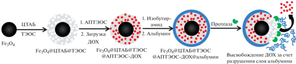
Среди биологических триггеров особый интерес представляет высвобождение лекарственного средства, которое обеспечивается действием фермента на биодеградацию защитного слоя на поверхности нанокомпозита. В работе [28] предложена конструкция биоразлагаемых материалов на основе МНЧ размером 30 нм, покрытых мезопористым слоем силикагеля. Поры этого слоя размером 3 нм были заполнены доксорубицином (35 мас. %). Для инкапсуляции действующего вещества было нанесено покрытие из альбумина11, связанного изобутирамидом (рис. 1). С помощью трехмерной клеточной модели и конфокальной визуализации показано, что эта белковая наноструктура разрушается под действием протеаз, высвобождая доксорубицин. Предложенная структура особенно интересна для разработки защитных слоев, использующих ферментативное разрушение в качестве триггера для высвобождения лекарств, и перспективна для высвобождения противоопухолевых препаратов и МРТ-диагностики.
Рис. 1.
Получение магнитных наночастиц для доставки доксорубицина и высвобождение препарата при разрушении защитного поверхностного слоя под действием фермента [28]. ЦТАБ – бромид цетилтриметиламмония, ТЭОС – тетраэтоксисилан, АПТЭОС – 3-аминопропилтриэтоксисилан, ДОХ – доксорубицин.
Для получения наноразмерных липидных везикул (липосом, керасом и магнитосом) используют свойство молекул фосфолипидов за счет своих поверхностно-активных свойств самопроизвольно образовывать в водной среде бислои, которые, в свою очередь, склонны к дальнейшему образованию полых пузырьковых структур (липосом). Это обеспечивает эффективное взаимодействие наноносителя с клеточной мембраной. Лекарственное средство может располагаться как в ядре липосомы, если оно водорастворимое, так и в ее оболочке, если лекарство жирорастворимое. Гораздо более высокую стабильность, чем у обычных липосом [29], демонстрируют керасомы. Магнитосомы [30–32] (рис. 2) – липосомы с дополнительно встроенными в структуру МНЧ – применяются не только в качестве магнитно-управляемого доставщика лекарств [31], но и позволяют контролировать свое распределение in vivo при помощи средств магнитно-резонансной визуализации. В работе [32] показано, что высокая устойчивость нагруженных доксорубицином магнитных керасом сохраняется в течение длительного времени.

Своеобразной оболочкой для МНЧ могут служить и другие объемные структуры, содержащие полости, размер которых достаточен для размещения МНЧ. Например, разработана [33] многофункциональная наноплатформа для мультимодальной визуализации и радиотерапии на основе наночастиц золота, стабилизированных поли(амидоамином) – дендримером 5-го порядка ветвления (G5), в которые добавлены НЧ оксида железа. Образующиеся стабилизированные дендримером МНЧ Fe3O4/Au имели диаметр около 100 нм, демонстрировали хорошую коллоидную стабильность, цитосовместимость и способность поглощать в ближней ИК-области.
Предложены и другие интересные структуры. Так, широкими потенциальными возможностями обладают многофункциональные материалы, описанные, в частности, в работе [34]: гантелеобразные (dumbbell) МНЧ Au–Fe3O4 с высоким значением магнитного момента, обеспечивающие визуализацию, характеризацию и определение на клеточном и субклеточном уровнях, а также полые МНЧ для доставки цисплатина и доксорубицина.
Следует отметить, что препараты, содержащие благородные металлы (Au, Ag, Pt, Pd) в виде инкорпорированных молекулярных форм, нанослоев на поверхности МНЧ или НЧ в их структуре, находят широкое применение в биомедицинских исследованиях [19, 34]: AuНЧ оптически активны и используются в оптической визуализации на основе светорассеяния, фотолюминисценции и гигантского комбинационного рассеяния (surface enhanced Raman scattering), а также поверхностного плазмонного резонанса в области 500–800 нм. Платина и палладий используются в основном в виде молекулярных форм.
Всесторонняя характеризация получаемых наноструктур – важнейшее условие последующего эффективного практического применения наноматериалов, поскольку их уникальные характеристики непосредственно зависят от структурных изменений на наноуровне, от конкретных размеров НЧ, степени их полидисперсности и морфологии (геометрии НЧ, размеров ядра и оболочек). В работах [8, 10, 35] детально рассмотрены преимущества и недостатки методов, применяемых для характеризации размеров, полидисперсности и геометрических характеристик ядра и оболочек НЧ.
БИОМЕДИЦИНСКИЕ ЗАДАЧИ, РЕШАЕМЫЕ С ИСПОЛЬЗОВАНИЕМ МАГНИТНЫХ НАНОЧАСТИЦ, И ИХ АНАЛИТИЧЕСКОЕ ОБЕСПЕЧЕНИЕ
Наиболее важным свойством МНЧ для биомедицинского применения является биосовместимость и способность к биологическому разложению, что позволяет транспортировать их по пути метаболизма железа [36]. Однако исследований, посвященных этой важнейшей проблеме, недостаточно, и взаимодействия в сыворотке крови человека из-за их сложности изучаются редко [37].
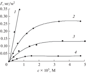
Ключевым вопросом при оценке клинической применимости препаратов является оценка их поведения в организме. В случае МНЧ как краткосрочные последствия применения (время циркуляции крови, биораспределение на суборганном и клеточном уровнях), так и долгосрочные (токсичность, скорость деградации частиц и выведения из организма) не вполне ясны. На эти факторы в экспериментах in vivo будет влиять, в частности, время циркуляции МНЧ в крови, которое может варьировать от нескольких минут до дней вследствие формирования на их поверхности белковой короны, которая неизбежно образуется за счет практически мгновенной сорбции белков из плазмы крови при контакте с НЧ [38]. Состав и структура белковой оболочки НЧ во многом зависит от физико-химических параметров НЧ: поверхности частицы, ее размера, формы, а также условий взаимодействия (источника белка, температуры, рН, длительности экспонирования) (рис. 3).
Рис. 3.
Изотермы адсорбции человеческого сывороточного альбумина на кремнеземе, модифицированном: 1 – полиэтиленимином; 2 – немодифицированном; 3 – 3-аминопропилтриэтоксисиланом; 4 – метилтриэтоксисиланом [39].
Классическое описание белковой короны подразумевает наличие “твердой” и “мягкой” корон [40]. Предполагают, что белки жесткой короны взаимодействуют непосредственно с поверхностью НЧ, а белки мягкой короны связываются с белками жесткой короны через слабые белок-белковые взаимодействия [41]; первоначальный состав короны формируется уже через 30 с инкубации НЧ [42]. Последствия таких нанобиовзаимодействий разнообразны и пока плохо предсказуемы. Они могут включать увеличение растворимости НЧ; иммунный ответ организма, направленный на устранение НЧ из кровотока; маскировку химических или биологических функций, специально придаваемых НЧ [38]. Например, установлено, что сорбция белков сыворотки крови на магнитных носителях (т.е. образование короны) препятствует агломерации МНЧ [43]. В детальных исследованиях, посвященных деградации МНЧ in vitro в имитируемых лизосомальных условиях [44, 45] и клеточных культурах [46, 47], установлены такие аспекты биотрансформации магнитных частиц, как высвобождение ионов металлов, участие комплекса метаболического белка железа в поглощении железа, постепенный переход частиц в форму ферритина и возможность синтеза частиц de novo из продуктов распада [48]. Вместе с тем комплексные исследования биотрансформации частиц железа в организмах проводятся редко [7]. В связи с этим выявление закономерностей формирования и последующего нанобиовзаимодействия белковой короны (с конкретным составом и структурой) для каждого типа НЧ является актуальной задачей.
Основными направлениями использования атомно- и масс-спектральных методов в биомедицинских исследованиях являются анализ биологических жидкостей и тканей, содержащих НЧ; определение растворенных и наноразмерных форм лекарственных препаратов в биообъектах (с разделением и без); определение размера НЧ, выявление характера распределения НЧ по размерам и его изменения при контакте с биологическими средами.
Общее содержание элементов, входящих в состав НЧ наноматериалов, чаще всего определяют методами МС-ИСП, реже – АЭС-ИСП или ЭТААС [49]; для решения этой задачи НЧ должны быть растворены в процессе минерализации матрицы или после ее щелочного или ферментативного расщепления.
Вследствие сложности биологических систем, содержащих НЧ, поведение частиц (например, скорость и степень деградации) в таких системах на начальных этапах часто исследуют в модельных экспериментах с использованием растворов, имитирующих биологические жидкости (белковые растворы, плазма или сыворотка крови), и на более простых биологических системах (инкубирование НЧ в присутствии клеточных культур, растительных и биологических матриц). При этом в любом случае необходима пробоподготовка, направленная на отделение матрицы, а также разделение растворенных и наноразмерных форм. Отделение наночастиц может быть выполнено путем магнитной сепарации, ультрафильтрации, с использованием методов капиллярного электрофореза или хроматографии. Например, для изучения изменений МНЧ при инкубации с белками в имитируемых физиологических условиях были исследованы возможности капиллярного электрофореза (КЭ) в сочетании с тандемной масс-спектрометрией (КЭ-МС/МС-ИСП) [50].
Спрос на быструю и эффективную методологию разделения и очистки продуктов, содержащих НЧ различной степени полидисперности, в известной степени может быть удовлетворен за счет ВЭЖХ в ее эксклюзионном варианте. Однако этот вид разделения обычно обладает низкой эффективностью разделения по причине медленной диффузии НЧ в жидкой фазе; возможны потери пробы из-за сорбционных взаимодействий НЧ с неподвижной фазой. В качестве альтернативы на примере ряда НЧ (золота, серебра и палладия, поверхностно-модифицированных органическими соединениями) показана возможность применения для разделения НЧ обращенно-фазовой ВЭЖХ (ОФ-ВЭЖХ) [51, 52]. Этот вариант ВЭЖХ отличается быстрым установлением динамического равновесия на колонке и, соответственно, высокой эффективностью разделения. По нашим предварительным данным, полученным при ОФ-ВЭЖХ-разделении гидрофильных НЧ магнетита, модифицированных цитрат-ионом (Fe3O4-Cit), этот вариант ОФ-ВЭЖХ вполне работоспособен. В сочетании со спектрофотометрическим детектированием в видимом диапазоне (400–450 нм), характерном для указанных НЧ, были получены хроматограммы, имеющие несколько воспроизводимых пиков, соответствующих фракциям, одновременно содержащим растворенное железо и МНЧ (подтверждено АЭС-ИСП-анализом фракций на содержание общего железа).
По сравнению с классическими вариантами МС-ИСП и оптической эмиссионной спектрометрией с ИСП, имеющих ряд инструментальных ограничений, SP-ICPMS позволяет получить гораздо больше информации, в частности, данные о концентрации частиц в единице объема анализируемого раствора и о распределении частиц по размерам. На основании этих данных можно судить об изменениях, происходящих с НЧ при контакте с биологической средой: происходит ли растворение НЧ, их агрегирование, накопление и какова скорость этих изменений. Однако даже для SP-ICPMS магнитные (т.е. железосодержащие) НЧ наиболее сложны. Несмотря на то, что метод позволяет определить число НЧ и их размеры [53], предел обнаружения железа в виде частиц существенно ухудшается из-за присутствия в организме эндогенного железа [54], главным образом в виде комплекса с ферритином и гемосидерином. Вследствие значимости метода SP-ICPMS для современных биомедицинских исследований мы рассмотрим его ниже более подробно.
К последним достижениям масс-спектрометрии относится также SС-ICPMS [55] – вариант, который позволяет анализировать отдельные клетки [56]. Отметим, что состав клетки намного сложнее, чем состав НЧ, содержание неорганических компонентов значительно ниже, они являются более крупными и хрупкими объектами, чем НЧ. Опубликованные к настоящему времени работы по SC-ICP-MS можно разделить на две основные категории [57]. Одна из них включает подходы, разработанные и используемые для мониторинга объектов, импортируемых в клетки (например, наночастиц, квантовых точек, металлсодержащих препаратов) [58, 59]. Ко второй, часто более сложной категории, относятся работы, посвященные определению эндогенных элементов в клетках [60–62]. В сложных экспериментах авторам работы [63] удалось выполнить определение в единичных клетках как эндогенных элементов (P, S, Fe, Cu и Zn), так и платины, введенной в виде цисплатина. Полученные результаты выявили различия в абсолютном количестве эндогенных элементов в клетках между различными типами клеток, что указывает на потенциал SC-ICP-MS в качестве инструмента “металлофингерпринтинга”.
Ключевым аспектом в развитии этого подхода является совершенствование способов введения клеточных суспензий в ИСП путем разработки специализированных систем ввода растворов (распылителей и распылительных камер), обеспечивающих целостность и максимальную эффективность транспортировки анализируемых клеток в источник.
МАСС-СПЕКТРОМЕТРИЯ В РЕЖИМЕ ДЕТЕКТИРОВАНИЯ ОДИНОЧНЫХ ЧАСТИЦ В БИОМЕДИЦИНСКИХ ИССЛЕДОВАНИЯХ
Один из наиболее перспективных современных методов описания и определения НЧ, SP-ICP-MS, был предложен как альтернатива микроскопическим исследованиям и вначале рассматривался как идеальный метод для атомной спектрометрии [64]. Основы и методология анализа в режиме SP-ICP-MS изложены в работах [65–68].
В качестве аналитов обычно выступают металлические НЧ на основе Au, Ag, Pt, НЧ, имеющие в своем составе оксиды – FexOy, CeO2, CuO, SiO2, TiO2, ZnO и “квантовые точки” (quantum dots) – CdSe, ZnS и т.д. [69].
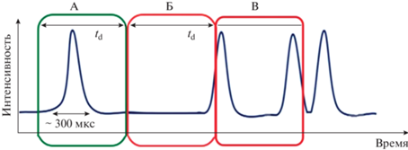
Анализ в режиме SP-ICP-MS выглядит следующим образом. Наночастицы в виде разбавленного коллоидного раствора, проходя через систему ввода пробы масс-спектрометра, попадают в плазму, где ионизируются, генерируя ионное облако. Правильно выбранное время интегрирования одиночного сигнала (dwell time, td) (от нескольких сотен микросекунд до нескольких миллисекунд) позволяет детектировать НЧ как одно событие (рис. 4). Количество событий, подсчитанных за время сбора данных, напрямую связано с количеством НЧ, поступающих в плазму. Далее количество событий может быть пересчитано в количество НЧ в растворе. Интенсивность каждого события пропорциональна количеству ионов в каждой отдельной НЧ и, следовательно, массе элемента в НЧ. Массу каждой НЧ можно преобразовать в ее размер, если известны состав, форма и плотность НЧ.
Рис. 4.
Корреляция между td и событиями, связанными с генерацией сигналов наночастиц (НЧ). А: Идеальное совпадение – одна НЧ регистрируется в течение выбранного td. Полученная интенсивность сигнала может быть использована для вычисления характеристик этой НЧ. Б: Неполная регистрация – сигнал НЧ регистрируется в двух временных отрезках. B: Регистрация события с двумя или несколькими НЧ, что может приводить к завышению размера НЧ.
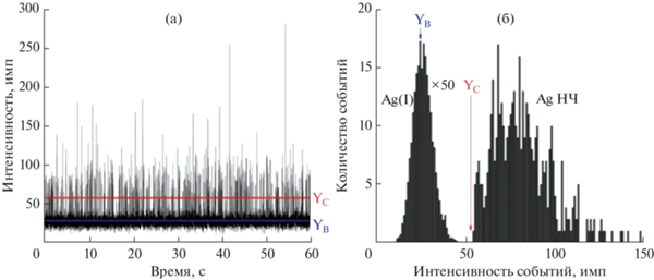
В SP-ICP-MS сигналы, генерируемые НЧ, записываются как временны́е развертки (рис. 5), которые состоят из “всплесков” (количество событий, связанных с НЧ), регистрируемых выше непрерывной базовой линии (фона). Полученные временны́е сканы можно обработать, построив зависимость количества событий от интенсивностей событий (или количество событий за фиксированное время сбора данных). Полученные таким образом гистограммы позволяют выделить область, связанную с информацией о растворенной форме элемента-аналита, и область, связанную с информацией о НЧ (рис. 5).
Рис. 5.
(а): Временной скан в режиме МС-ИСП одиночных частиц коллоидного раствора, содержащего НЧ Ag (100 нм) и 1 нг/мл растворенного серебра. (б): Гистограмма, построенная с учетом данных временного скана (а). Yb – среднее значение фонового сигнала, Yc – интенсивность, выше которой сигналы приписываются наночастицам [70].
В зависимости от выбранного td, события, связанные с НЧ, могут быть представлены в виде пиков, построенных с использованием одного (td в миллисекундном диапазоне 3–10 мс) или нескольких значений (td в микросекундном диапазоне 10–200 мкс) аналитических сигналов. Поскольку длительность события НЧ находится в диапазоне 300–1000 мкс [71], сигналы, полученные при разных значениях td, будут отличаться друг от друга (рис. 6).

Как отмечалось выше, временны́е сканы могут быть обработаны путем построения гистограмм зависимости количества событий от интенсивностей событий (рис. 5). Для отделения фоновых событий и событий с НЧ используют критерии отбора, выбор которых влияет на предел определения размера НЧ (ПрО размера НЧ). Обычно используют критерий 3δ, основанный на стандартном отклонении базовой линии [68]. Показано [72], что появления ложных результатов позволяет избежать использование критерия 5δ. Все предложенные алгоритмы могут быть реализованы пользователями, экспортирующими необработанные данные в собственные программы и электронные таблицы.
В методе SP-ICP-MS выделяют ПрО размера НЧ (LODsize), минимальную концентрацию НЧ (смин НЧ или LODНЧ) и минимальную массу регистрируемой НЧ (mмин НЧ).
ПрО размера НЧ в SP-ICP-MS определяется элементным составом, плотностью и формой НЧ и зависит от чувствительности спектрометра, а также выбранных условий измерений, особенно от td и от значений фона. ПрО размера НЧ, полученные для чистых водных коллоидных растворов, недостижимы для реальных образцов. Важную роль при нахождении ПрО размера НЧ играют изобарические и полиатомные влияния, которые обычно присутствуют в сложных матрицах; все возможные спектральные и неспектральные влияния должны быть выявлены, минимизированы и учтены. В качестве ориентира по ПрО размера НЧ, получаемым методом квадрупольной МС-ИСП, можно использовать теоретические расчеты для 40 различных синтезированных НЧ на фоне деионизованной воды [73]. В табл. 1 приведена информация о ПрО размера для наиболее хорошо изученных НЧ.
Таблица 1.
| Элемент | Изотоп | Состав НЧ | ПрО размера НЧ, нм | |
|---|---|---|---|---|
| td = 5 мс | td = 100 мкс | |||
| Ag | 107 | Ag | 17.4 | 12.3 |
| Au | 197 | Au | 12.7 | 9.0 |
| Pt | 195 | Pt | 5.3 | 3.7 |
| Si | 29 | SiO2 | 148.0 | 105.0 |
| Ti | 47 | TiO2 | 41.4 | 29.3 |
| Fe | 56 | Fe | 36** | |
Минимальная концентрация смин НЧ определяется минимальным количеством НЧ, которое достигает системы детектирования. Данное значение, как и число НЧ в 1 мл, рассчитывается по формуле (1):
где DНЧ − общее количество событий, связанных с НЧ; D – общее количество событий за выбранный промежуток времени измерения; η – эффективность распыления; q – скорость подачи раствора, мл/с; td – время накопления сигнала, с.Содержание НЧ в 1 мл зависит от эффективности транспортировки аэрозоля ƞ (количество растворенной формы аналита или НЧ, которое достигает плазмы; формула (2)) и доли частиц в растворе с размером выше ПрО размера НЧ. Допускают, что максимальное число событий с НЧ в холостых растворах не должно превышать 10 НЧ за минуту анализа, а минимальное количество событий НЧ в анализируемых растворах должно быть не менее 100 в минуту [75]. Обычно Смин НЧ имеет порядок 103 НЧ/мл [76, 77].
(2)
$\eta = \frac{{d{\text{\;}}{\kern 1pt} S{\kern 1pt} {{f}_{{\text{a}}}}{\text{\;}}{\kern 1pt} {{\pi \;}}{\kern 1pt} {{\rho }}}}{{6{\text{\;}}\left( {{{I}_{{{\text{НЧ}}}}} - {{I}_{{\text{ф}}}}} \right){{t}_{{\text{d}}}}~q}},$Величина mмин НЧ зависит от размера частиц и чувствительности спектрометра [78]. Массу НЧ определяет плотность вещества и доля аналита в НЧ. mмин НЧ, как и масса НЧ, рассчитывается по формуле (3):
где ρ – плотность вещества, из которого состоит НЧ; fa – массовое распределение элемента-аналита в НЧ; d – диаметр НЧ, нм. При расчете массы делается допущение, что НЧ имеет сферическую форму.Для режима МС-ИСП одиночных частиц характерны те же спектральные и матричные влияния, что и для обычного анализа методом МС-ИСП. Хорошо известны полиатомные влияния 40Ar16O+ на изотоп 56Fe+ и 40Ar16OH+ на изотоп 57Fe+. Минимизировать влияния аргона на изотоп 56Fe+ при работе с МНЧ можно за счет использования реакционной ячейки c водородом [75, 79], с аммиаком [75], а также коллизионной ячейки, наполненной гелием, или за счет высокого разрешения масс-спектрометра, например магнитно-секторного [80]. На рис. 5 представлен спектр изотопа 56Fe и 40Ar16O+, полученный нами на МС-ИСП-спектрометре Element XR (Finnigan Mat, Германия) в среднем разрешении. Видно, что пики 40Ar16O+ и 56Fe+ хорошо разрешаются (рис. 7).

Для получения количественной информации о размере и концентрации НЧ используют стандартные образцы НЧ разных размеров, имеющими тот же элементный состав, ту же геометрию и плотность, что и целевая НЧ. Однако нехватка монодисперсных, хорошо охарактеризованных и стабильных стандартов НЧ ограничивает применимость этого подхода. Можно использовать “home made” НЧ при условии их тщательного описания другими методами анализа, в частности с привлечением различных вариантов электронной микроскопии (просвечивающая, сканирующая), а также метода динамического рассеяния света. Рабочие коллоидные растворы должны быть стабильны при длительном хранении.
Кроме обычных требований к анализируемой пробе по составу и минерализации со стороны метода МС-ИСП, важным условием подготовки проб, содержащих НЧ, является исключение изменения и/или потери НЧ. Необходимо учитывать устойчивость суспензий НЧ в процессе хранения, обращения, пробоподготовки, поскольку изменение состава дисперсионной среды, разбавление, взаимодействие с материалом контейнера, условия и время хранения могут изменить состояние поверхностного слоя или размер НЧ и вызвать агрегацию. Принципиально важно учитывать возможность изменения состояния НЧ при фильтровании (в результате взаимодействия с материалом мембраны), а также вероятность взаимопревращений растворенных форм и НЧ при экстракции (извлечении), разложении или хранении.
Прямой анализ жидких образцов без пробоподготовки возможен только для наиболее простых систем; в этом случае чаще всего необходимо разбавление [81]. При этом для количественного МС-ИСП-анализа в режиме одиночных частиц необходимо скорректировать коэффициент разбавления таким образом, чтобы уменьшить вероятность обнаружения более чем одной НЧ в пределах выбранного td (исключить событие с несколькими НЧ), т.е. определить верхнюю границу содержаний НЧ в растворе. В то же время количество НЧ должно быть достаточным для удовлетворительных расчетов (чтобы уменьшить случайную погрешность, определяемую статистикой Пуассона), т.е. определить нижнюю границу содержаний НЧ в растворе [71]. Многократные разбавления пробы помогают снизить влияние матрицы, например при определении НЧ оксида железа в присутствии растворенного железа [82], или снизить влияния растворимого железа, входящего в состав гемоглобина [83]. Данные об определении НЧ золота и серебра различных размеров в растворах, полученных разбавлением биологических жидкостей водой, приведены в работах [84–86].
Анализ более сложных объектов может быть проведен после ферментативного или щелочного гидролиза, а также с привлечением лазерной абляции (ЛА). Ферментативный гидролиз [86–88] позволяет разрушить структуру матрицы, при этом фермент должен соответствовать типу образца. Например, протеиназы выбирают для гидролиза мышечной ткани, липазы для жировой ткани и т.д. Для проведения ферментативного гидролиза необходимо создать условия, способствующие максимальной активности фермента (оптимальный pH, присутствие Mg или Ca и аденозинтрифосфата). Широко используемым ферментом является протеиназа К, проявляющая активность в диапазоне рН 7.5–12.0 при 37°C [89]. Протеиназа К является Са-зависимым ферментом, поэтому для гидролиза биологических матриц используют реакционную смесь, содержащую 1–5 М раствор Ca2+, часто в сочетании с 1%-ным раствором Тритона X-100 и/или 0.5%-ным раствором додецилсульфата натрия [90, 91].
Для подготовки биологических тканей для МС-ИСП-анализа в режиме одиночных частиц используют также щелочной гидролиз в присутствии гидроксида тетраметиламмония (TMAOH) при рН > 14. Схема щелочного гидролиза включает добавление TMAOH до концентрации 10–20% (масса/объем) и объема 20–50 мкл на 1 мг биологической ткани, в результате чего конечный объем составляет 0.25–10 мл. Исключением являются случаи, например, c клеточными культурами, где образцы состоят из небольшого количества биомассы, поэтому TMAOH находится в большом избытке по сравнению с ней [92, 93]. Перед анализом полученные гидролизаты разбавляют сверхчистой водой таким образом, чтобы конечная концентрация ТМАOН составляла 1%.
Сравнивая щелочной (на основе TMAOH) и ферментативный гидролиз (с использованием протеиназы K), авторы отмечают, что если исследованные образцы содержат большое количество липидов, то ТМАOН более предпочтителен, так как протеиназа К не расщепляет жировые ткани [94, 95]. Вместе с тем можно предположить, что жесткие условия щелочного гидролиза приведут к изменению состояния НЧ и их оболочек. Имеющиеся данные о влиянии условий на размеры частиц противоречивы, особенно для наиболее мелких из детектируемых НЧ, что обусловлено, скорее всего, значительной погрешностью определения их размера вблизи ПрО [84, 93, 96–98].
Перспективным подходом для получения дополнительной информации о смесях НЧ представляется онлайн сочетание SP-ICP-MS с методами разделения. Основной проблемой этого сочетания является то, что SP-ICP-MS дискретно детектирует индивидуальные НЧ, тогда как методы разделения обеспечивают локальное концентрирование аналитов определенного вида (в виде пика), которые потом совместно элюируются с колонки/капилляра. Кроме того, в методах разделения используют среды (в основном органические соединения), которые при введении в плазму могут приводить к матричным спектральным влияниям. По этой причине совместное использование методов разделения и SP-ICP-MS требует тщательной оптимизации условий, которые должны обеспечить возможность детектирования отдельных частиц, отсутствие превращений НЧ в процессе выделения, а также исключить влияние органического буфера на детектирование.
Как следует из детального обзора [77], число комбинированных методов SP-ICP-MS-определения НЧ невелико. Описаны онлайн сочетания SP-ICP-MS с методами гидродинамической хроматографии, капиллярного электрофореза, фракционирования в потоке. Большинство методов для стабилизации частиц использует ПАВ (додецилсульфат натрия). Работы выполнены в основном на модельных смесях золотых и серебряных НЧ. Отдельные работы посвящены Pt-содержащим препаратам и разделению частиц и их растворенных форм [99, 100]. В работе [99] поведение НЧ на основе оксидов железа контролировали с помощью ВЭЖХ-МС-ИСП с детектором, работающим в режиме коллизионной ячейки для устранения 40Ar16O+ и 40Ar16OH+ при измерении 56Fe+ одновременно с 195Pt+.
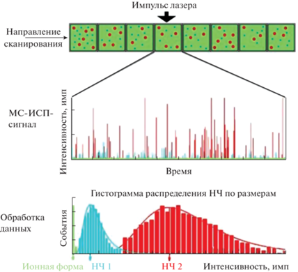
В SP-ICP-MS для анализа образцов, содержащих НЧ, кроме прямого ввода коллоидного раствора в масс-спектрометр, можно использовать лазерную абляцию. Сочетание масс-спектрометра и системы ЛА является оптимальным для биовизуализации, поскольку обеспечивает возможность многоэлементного анализа, характеризуется низкими пределами определения элементов (на уровне мкг/г) и высоким пространственным разрешением (до уровня мкм) [101].
При ЛА-SP-ICP-MS-анализе биологической матрицы, содержащей растворенную форму и НЧ элемента-аналита, формируется комбинированный сигнал, состоящий из непрерывного сигнала растворенной формы и импульсов НЧ (рис. 8) [102]. Далее обработка сигналов идет как в обычном режиме SP-ICP-MS. При этом могут возникнуть трудности, обусловленные высоким содержанием растворенной формы элемента, которое мешает определению НЧ. Кроме того, локализация НЧ на одном участке биоматериала может привести к одновременному обнаружению нескольких НЧ. В отличие от анализа растворов методом SP-ICP-MS, где события можно разрешить, например, за счет разбавления, в ЛА-SP-ICP-MS меньше возможностей при выборе и варьировании условий эксперимента.
Рис. 8.
Этапы получения и обработки аналитического сигнала методом ЛА-МС-ИСП в режиме детектирования одиночных частиц [101].
Отсутствие сертифицированных эталонных материалов для приложений биовизуализации является большой проблемой как для ЛА-SP-ICP-MS, так и для SP-ICP-MS в целом. В настоящее время большое внимание уделяется поиску и разработке стандартных образцов, в том числе на основе биологических матриц. Так, для градуировки предложены стандарты на полимерной основе (желатине) [103].
Перспективным подходом к анализу сложных биологических матриц представляется сочетание изотопного разбавления и SP-ICP-MS [104]. Так, предложена методика определения размеров НЧ Ag (от 30 до 80 нм) с добавлением изотопа 109Ag+. В данном случае изотопное разбавление позволяет компенсировать подавление сигнала элемента-аналита в присутствии высоких концентраций NaCl.
* * *
Большой интерес к функционализированным наноматериалам с заданными свойствами для разработки терапевтических средств и других бионанотехнологических приложений обусловлен широкими возможностями, которые предоставляют такие материалы для решения задач диагностики и целевой доставки лекарственных препаратов. Отличительной чертой подходов, реализуемых при решении задач с использованием наноразмерных материалов (в том числе обладающих магнитными свойствами), является вовлечение в исследования большого числа инструментальных методов. Это обусловлено тем, что для рационального проектирования микро- и наночастиц с контролируемой скоростью разложения и низкой токсичностью in vivo необходима их полная характеризация и выявление фундаментальных закономерностей, обусловливающих скорость биотрансформации магнитного материала и его выведения из организма в зависимости от дозы частиц, их гидродинамического размера, внутренней структуры и природы внешнего покрытия. Значимое место в исследованиях частиц, входящих в состав наноматериалов, занимают многоэлементные спектрометрические методы, особенно масс-спектрометрия с индуктивно связанной плазмой. Различные ее варианты позволяют всесторонне охарактеризовать поведение нанопрепаратов в биосистемах, предоставляя информацию не только о числе и размерах частиц, но и об их растворенных формах в биологических жидкостях и тканях. Сочетание же МС-детектирования с методами разделения характеризует распределение элементов и их форм даже в отдельных клетках. Вместе с тем новые подходы к анализу сложных биологических сред пока находятся в стадии разработки, требуют инновационных инструментальных и методических решений и создания надежной методической базы для проведения комплексных аналитических и биомедицинских исследований.
Список литературы
Nochehdehi A.R., Thomas S., Sadri M., Afghahi S.S.S., Mehdi Hadavi S.M. Iron oxide biomagnetic nanoparticles (IO-BMNPs); synthesis, characterization and biomedical application − A review // J. Nanomed. Nanotechnol. 2017. V. 8. № 1. P. 423. https://doi.org/10.4172/2157-7439.1000423
Королев Д.В. Разработка препаратов для тераностики и направленной доставки кардиопротективных субстанций на основе кремнеземных и магнитных наночастиц Дис. … док. хим. наук. Санкт-Петербург: Национальный медицинский исследовательский центр им. В.А. Алмазова Министерства здравоохранения РФ, 2019. 384 с.
Sandler S.E., Fellows B., Mefford O.T. Best practices for characterization of magnetic nanoparticles for biomedical applications // Anal. Chem. 2019. V. 91. P. 14159. https://doi.org/10.1021/acs.analchem.9b03518
Ganta S., Devalapally H., Shahiwala A., Amiji M. A review of stimuli-responsive nanocarriers for drug and gene delivery// J. Control. Release. 2008. V. 126. № 3. P. 187. https://doi.org/10.1016/j.jconrel.2007.12.017
Nanoparticles for Drug Delivery / Eds. Joshy K.S., Sabu Thomas, Vijay Kumar Thakur. Springer Nature Singapore Pte Ltd., 2021. https://doi.org/10.1007/978-981-16-2119-2
Xu C., Sun S. New forms of superparamagnetic nanoparticles for biomedical applications // Adv. Drug Deliv. Rev. 2013. V. 65. № 5. P. 732. https://doi.org/10.1016/j.addr.2012.10.008
Zelepukin I.V., Yaremenko A.V., Ivanov I.N., Yuryev M.V., Cherkasov V.R., Deyev S.M., Nikitin P.I., Nikitin M.P. Long-term fate of magnetic particles in mice: A comprehensive study // ACS Nano. 2021. V. 15. P. 11341. https://doi.org/10.1021/acsnano.1c00687
Handbook of Bioanalytics / Eds. Buszewski B., Baranowska I. Springer Nature Switzerland AG, 2022. P. 1091. https://doi.org/10.1007/978-3-030-95660-8
ICPMS and trace element analysis as tools for better understanding medical conditions / Comprehensive Analytical Chemistry / Eds. Aurélio M., Arruda Z., Jemmyson Romário de Jesu, 2022. V. 97. P. 2. https:// www.sciencedirect.com/science/journal/0166526X/ 97/ supp/Cю
Hurley K.R., Ring H.L., Kang H., Klein N.D., Haynes C.L. Characterization of magnetic nanoparticles in biological matrices // Anal. Chem. 2015. V. 87. P. 11611. https://doi.org/10.1021/acs.analchem.5b02229
Anselmo A.C., Mitragotri S. Nanoparticles in the clinic: An update // Bioeng. Transl. Med. 2019. V. 4. № 3. Article e10143. https://doi.org/10.1002/btm2.10143
Kharisov B.I., Rasika Dias H.V., Kharissova O.V., Vazquez A., Pena Y., Gomez I. Solubilization, dispersionand stabilization of magnetic nanoparticles in water and non-aqueous solvents: Recent trends // RSCAdv. 2014. V. 4. № 85. P. 45354. https://doi.org/10.1039/C4RA06902A
Xie L., Jiang R., Zhu F., Liu H., Ouyang G. Application of functionalized magnetic nanoparticles in samplepr eparation // Anal. Bioanal. Chem. 2014. V. 406. № 2. P. 377. https://doi.org/10.1007/s00216-013-7302-6
Пряжников Д.В., Кубракова И.В. Магнитные наноразмерные материалы с модифицированной поверхностью: получение и исследование структуры, состава и свойств // Журн. аналит. химии. 2021. Т. 76. № 6. С. 496. https://doi.org/10.31857/S0044450221060098 (Pryazhnikov D.V., Kubrakova I.V. Surface-modified magnetic nanoscale materials: Preparation and study of their structure, composition, and properties // J. Anal. Chem. 2021. Т. 76. № 6. P. 685.)
Kudr J., Haddad Y., Richtera L., Heger Z., Cernak M., Adam V., Zitka O. Magnetic nanoparticles: From design and synthesis to real world applications // Nanomaterials. 2017. V. 7. № 9. P. 243. https://doi.org/10.3390/nano7090243
Pryazhnikov D.V., Kubrakova I.V., Kiseleva M.S, Martynov L.Yu., Koshcheeva I.Ya. Preparation and structural characterization of nanosized magnetic solid-phase extractants // Mendeleev Commun. 2014. V. 24. № 2. P. 130. https://doi.org/10.1016/j.mencom.2014.03.023
Кубракова И.В., Пряжников Д.В. Микроволновый синтез наноразмерных магнитных сорбентов // Журн. аналит. химии. 2021. Т. 76. № 1. С. 20. https://doi.org/10.31857/S0044450221010047 (Kubrakova I.V., Pryazhnikov D.V. Microwave-assisted synthesis of nanosized magnetic sorbents // J. Anal. Chem. 2021. Т. 76. № 1. С. 15.)
Mekseriwattana W., Srisuk S., Kriangsaksri R., Niamsiri N., Prapainop K. The impact of serum proteins and surface chemistry on magnetic nanoparticle colloidal stability and cellular uptake in breast cancer cells // AAPS PharmSciTech. 2019. V. 20. P. 55. https://doi.org/10.1208/s12249-018-1275-x
Timerbaev A.R. Analytical methodology for developing nanomaterials designed for magnetically-guided delivery of platinum anticancer drugs // Talanta. 2022. V. 243. Article 123371. https://doi.org/10.1016/j.talanta.2022.123371
Nigam S., Chandra S., Newgreen D.F., Bahadur D., Chen Q. Poly (ethylene glycol)-modified PAMAM-Fe3O4-doxorubicin triads with the potential for improved therapeutic efficacy: generation-dependent increased drug loading and retention at neutral pH and increased release at acidic pH // Langmuir. 2014. V. 30. P. 1004. https://doi.org/10.1021/la404246h
Gaihre B., Khil M.S., Lee D.R., Kim H.Y. Gelatin-coated magnetic iron oxide nanoparticles as carrier system: Drug loading and in vitro drug release study // Int. J. Pharm. 2009. V. 365. P. 180. https://doi.org/10.1016/j.ijpharm.2008.08.020
Lee J.E., Lee D.J., Lee N., Kim B.H., Choi S.H., Hyeon T. Multifunctional mesoporous silica nanocomposite nanoparticles for pH controlled drug release and dual modal imaging // J. Mater. Chem. 2011. V. 21. P. 16869. https://doi.org/10.1039/C1JM11869B
N’Guyen T.T.T., Duong H.T.T., Basuki J., Montembault V., Pascual S., Guibert C., Fresnais J., Boyer C., Whittaker M.R., Davis T.P., Fontaine L. Functional iron oxide magnetic nanoparticles with hyperthermia-induced drug release ability by using a combination of orthogonal click reactions // Angew. Chem. Int. Ed. 2013. V. 52. P. 14152. https://doi.org/10.1002/anie.201306724
Yuan L., Tang Q., Yang D., Zhang J.Z., Zhang F., Hu J. Preparation of pH-responsivemesoporous silica nanoparticles and their application in controlled drug delivery // J. Phys. Chem. C. 2011. V. 115. P. 9926. https://doi.org/10.1021/jp201053d
Shen J., He Q., Gao Y., Shi J., Li Y. Mesoporous silica nanoparticles loading doxorubicin reverse multidrug resistance: Performance and mechanism // Nanoscale. 2011. V. 3. P. 4314. https://doi.org/10.1039/c1nr10580a
Пряжников Д.В., Ефанова О.О., Киселева М.С., Кубракова И.В. Микроволновый синтез наноразмерных материалов типа “ядро–оболочка” на основе магнетита, функционализированного золотом и доксорубицином // Российские нанотехнологии. 2017. Т. 12. № 3–4. С. 69. doi: 10.1134/S1995078017020094 (Pryazhnikov D.V., Efanova O.O., Kiseleva M.S., Kubrakova I.V. Microwave synthesis of core–shell nanosize materials on the basis of magnetite functionalized with gold and doxorubicine // Nanotechnologies in Russia. 2017. V. 12. № 3–4. P. 199.)
Liu J., Detrembleur C., De Pauw-Gillet M.-C., Mornet S., Vander Elst L., Laurent S., Jérôme C., Duguet E. Heat-triggered drug release systems based on mesoporous silica nanoparticles filled with a maghemite core and phase-change molecules as gatekeepers // J. Mater. Chem. B. 2014. V. 2. P. 59. https://doi.org/10.1039/c3tb21229g
Ménard M., Meyer F., Affolter-Zbaraszczuk C., Rabineau M., Adam A., Duenas Ramirez P., Bégin-Colin S., Mertz D. Design of hybrid protein-coated magnetic core mesoporous silica shell nanocomposites for MRI and drug release assessed in a 3D tumor cell model // Nanotechnology. 2019. V. 30. № 17. Article 174001. https://doi.org/10.1088/1361-6528/aafe1c
Wang Y., Wang B., Liao H., Song X., Wu H., Wang H., Shen H., Ma X., Tan M. Liposomal nanohybrid cerasomes for mitochondria-targeted drug delivery // J. Mater. Chem. B. 2015. V. 3. P. 7291. https://doi.org/10.1039/c5tb01197c
Jin Y., Yue X., Zhang Q., Wu X., Cao Z., Dai Z. Cerasomal doxorubicin with long-term storage stability and controllable sustained release // Acta Biomaterialia. 2012. V. 8. P. 3372. https://doi.org/10.1016/j.actbio.2012.05.022
Bixner O., Reimhult E. Controlled magnetosomes: Embedding of magnetic nanoparticles into membranes of monodisperse lipid vesicles // J. Colloid Interface Sci. 2016. V. 466. P. 62. https://doi.org/10.1016/j.jcis.2015.11.071
Pryazhnikov D.V., Efanova O.O., Kubrakova I.V. Cerasomes containing magnetic nanoparticles: Synthesis and gel-filtration chromatographic characterization // Mendeleev Commun. 2019. V. 29. № 2. P. 226. https://doi.org/10.1016/j.mencom.2019.03.038
Lu S., Li X., Zhang J., Peng C., Shen M., Shi X. Dendrimer-stabilized gold nanoflowers embedded with ultrasmall iron oxide nanoparticles for multimode imaging-guided combination therapy of tumors // Adv Sci (Weinh). 2018. V. 5. № 12. Article 1801612. https://doi.org/10.1002/advs.201801612
Xu C., Sun S. New forms of superparamagnetic nanoparticles for biomedical applications // Adv. Drug Deliv. Rev. 2013. V. 65. P. 732. https://doi.org/10.1016/j.addr.2012.10.008
Rios A., Zougagh M. Recent advances in magnetic nanomaterials for improving analytical processes // Trends Anal. Chem. 2016. V. 84 A. P. 72. https://doi.org/10.1016/j.trac.2016.03.001
Alshehri S., Imam S.S., Rizwanullah M., Akhter S., Mahdi W., Kazi M., Ahmad J. Progress of cancer nanotechnology as diagnostics, therapeutics, and theranostics nanomedicine: preclinical promise and translational challenges // Pharmaceutics. 2021. V. 13. P. 24. https://doi.org/10.3390/pharmaceutics13010024
Timerbaev A.R. How well can we characterize human serum transformations of magnetic nanoparticles // Analyst. 2020. V. 145. P. 1103. https://doi.org/10.1039/C9AN01920K
Ke P.C., Lin S., Parak W.J., Davis T.P., Caruso F. Decade of the Protein Corona // ACS Nano. 2017. T. 11. № 12. P. 11773. https://doi.org/10.1021/acsnano.7b08008
Куликова Г.А., Парфенюк Е.В. Поверхностные свойства модифицированных наноразмерных кремнеземов и их влияние на иммобилизацию человеческого сывороточного альбумина // Физикохимия поверхности и защита материалов. 2010. Т. 46. № 5. С. 473. (Kulikova G.A., Parfenyuk E.V. Surface properties of modified nanosized silica and their influence on human serum albumin immobilization // Prot. Met. Phys. Chem. Surf. 2010. Т. 46. № 5. P. 546.)
Winzen S., Schoettler S., Baier G., Rosenauer C., Mailaender V., Landfester K., Mohr K. Complementary analysis of the hard and soft protein corona: sample preparation critically effects corona composition // Nanoscale. 2015. T. 7. № 7. C. 2992. https://doi.org/10.1039/c4nr05982d
Lundqvist M., Stigler J., Elia G., Lynch I., Cedervall T., Dawson K.A. Nanoparticle size and surface properties determine the protein corona with possible implications for biological impacts // Proc. Natl. Acad. Sci. U.S.A. 2008. V. 105. № 38. P. 14265. https://doi.org/10.1073/pnas.0805135105
Tenzer S., Docter D., Kuharev J., Musyanovych A., Fetz V., Hecht R., Schlenk F., Fischer D., Kiouptsi K., Reinhardt C., Landfester K., Schild H., Maskos M., Knauer S.K., Stauber R.H. Rapid formation of plasma protein corona critically affects nanoparticle pathophysiology // Nat. Nanotechnol. 2013. T. 8. № 10. P. 772. https://doi.org/10.1038/nnano.2013.181
Sikorski J., Matczuk M., Kaminska A., Kruszewska J., Trzaskowski M., Timerbaev A.R., Jarosz M. Protein-mediated transformations of superparamagnetic nanoparticles evidenced by single-particle inductively coupled plasma tandem mass spectrometry: A disaggregation phenomenon // Int. J. Mol. Sci. 2022. V. 23. P. 1088. https://doi.org/10.3390/ijms23031088
Rabel M., Warncke P., Grüttner C., Bergemann C., Kurland H.-D., Müller R., Dugandzic V., Thamm J., Muller R., Popp J., Cialla-May D., Fischer D. Simulation of the long-term fate of superparamagnetic iron oxide-based nanoparticles using simulated biological fluids // Nanomedicine. 2019. V. 14. P. 1681. https://doi.org/10.2217/nnm-2018-0382
Gutiérrez L., Romero S., Da Silva G.B., Costo R., Vargas M.D., Ronconi C.M., Serna C.J., Veintemillas-Verdaguer S., Del Puerto Morales M. Degradation of magnetic nanoparticles mimicking lysosomal conditions followed by AC susceptibility // Biomed. Tech. 2015. V. 60. P. 417. https://doi.org/10.1515/bmt-2015-0043
Rojas J.M., Gavilán H., Del Dedo V., Lorente-Sorolla E., Sanz-Ortega L., Da Silva G.B., Costo R., Perez-Yagüe S., Talelli M., Marciello M., Morales M.P., Barber D.F., Gutiérrez L. Time course assessment of the aggregation and metabolization of magnetic nanoparticles // Acta Biomater. 2017. V. 58. P. 181. https://doi.org/10.1016/j.actbio.2017.05.047
Yurenya A., Nikitin A., Garanina A., Gabbasov R., Polikarpov M., Cherepanov V., Chuev M., Majouga A., Panchenko V. Synthesis and mössbauer study of 57Fe-based nanoparticles biodegradation in living cells // J. Magn. Magn. Mater. 2019. V. 474. P. 337. https://doi.org/10.1016/j.jmmm.2018.11.040
Mazuel F., Espinosa A., Luciani N., Reffay M., Le Borgne R., Motte L., Desboeufs K., Michel A., Pellegrino T., Lalatonne Y., Wilhelm C. Massive intracellular biodegradation of iron oxide nanoparticles evidenced magnetically at single-endosome and tissue levels // ACS Nano. 2016. V. 10. P. 7627. https://doi.org/10.1021/acsnano.6b02876
Седых Э.М., Дементьева О.В., Карцева М.Е., Румянцева Т.Б., Тунян А.А., Банных Л.Н., Громяк И.Н., Рудой В.М. Возможности методов атомной спектроскопии при анализе наночастиц на основе золота и серебра в синтезируемых золях и биологических объектах // Журн. аналит. химии. 2016. Т. 71. № 1. С. 65. doi: 10.7868/S0044450216010138 (Sedykh E.M., Dement’eva O.V., Kartseva M.E., Roumyantseva T.B., Tunyan A.A., Bannykh L.N., Gromyak I.N., Rudoy V.M. Possibilities of atomic spectroscopy in the analysis of gold-and silver-based nanoparticles in synthesized sols and biological samples // J. Anal. Chem. 2016. V. 71. P. 62.)
Kruszewska J., Sikorski J., Samsonowicz-Górski J., Matczuk M. A CE-ICP-MS/MS method for the determination of superparamagnetic iron oxide nanoparticles under simulated physiological conditions // Anal. Bioanal. Chem. 2020. V. 412. P. 8145. https://doi.org/10.1007/s00216-020-02948-3
Choi M.M.F., Douglas A.D., Murray R.W. Ion-pair chromatographic separation of water-soluble gold monolayer-protected clusters // Anal. Chem. 2006. V. 78. № 8. P. 2779. https://doi.org/10.1021/ac052167m
Zhang Y., Shuang S., Dong C., Lo C.K., Paau M.C., Choi M.M.F. Application of HPLC and MALDI-TOF MS for studying As-synthesized ligand-protected gold nanoclusters products // Anal. Chem. 2009. V. 81. № 4. P. 1676. https://doi.org/10.1021/ac8026349
Pace H.E., Rogers N.J., Jarolimek C., Coleman V.A., Higgins C.P., Ranville J.F. Correction to determining transport efficiency for the purpose of counting and sizing nanoparticles via single particle inductively coupled plasma mass spectrometry // Anal. Chem. 2012. V. 84. P. 4633. https://doi.org/10.1021/ac201952t
Levy M., Luciani N., Alloyeau D., Elgrabli D., Deveaux V., Pechoux C., Chat S., Wang G., Vats N., Gendron F., Factor C., Lotersztajn S., Luciani A., Wilhelm C., Gazeau F. Long term in vivo biotransformation of iron oxide nanoparticles // Biomaterials. 2011. V. 32. P. 3988. https://doi.org/10.1016/j.biomaterials.2011.02.031
Montes-Bayón M., Corte-Rodríguez M., Álvarez-Fernández García R., Severo Fagundes J. Biomedical analysis by ICP-MS: A focus on single cell strategies / Comprehensive Analytical Chemistry. 2022. V. 97. Ch. 4. P. 109. https://doi.org/10.1016/bs.coac.2022.03.002
Ivask A., Mitchell A.J., Malysheva A., Voelcker N.H., Lombi E. Methodologies and approaches for the analysis of cell nanoparticle interactions // Nanomed. Nanobiotechnol. 2018. V. 10. Article e1486. https://doi.org/10.1002/wnan.1486
Yu X., He M., Chen B., Hu B. Recent advances in single-cell analysis by inductively coupled plasma-mass spectrometry: A review // Anal. Chim. Acta. 2020. V. 1137. P. 191. https://doi.org/10.1016/j.aca.2020.07.041
Sun Q.-X., Wei X., Zhang S.-Q., Chen M.-L., Yang T., Wang J.-H. Single cell analysis for elucidating cellular uptake and transport of cobalt curcumin complex with detection by time-resolved ICPMS // Anal. Chim. Acta. 2019. V. 1066. P. 13. https://doi.org/10.1016/j.aca.2019.03.062
García R.A.-F., Corte-Rodríguez M., Macke M., LeBlanc K., Mester Z., Montes-Bayon M., Bettmer J. Addressing the presence of biogenic selenium nanoparticles in yeast cells: Analytical strategies based on ICP-TQ-MS // Analyst. 2020. V. 145. P. 1457. https://doi.org/10.1039/c9an01565e
Cao Y., Feng J., Tang L., Yu C., Mo G., Deng B. A highly efficient introduction system for single cell-ICP-MS and its application to detection of copper in single human red blood cells // Talanta. 2020. V. 206. Article 120174. https://doi.org/10.1016/j.talanta.2019.120174
Amor M., Tharaud M., Gelabert A., Komeili A. Single-cell determination of iron content in magnetotactic bacteria: Implications for the iron biogeochemical cycle // Environ. Microbiol. 2020. V. 22. P. 823. https://doi.org/10.1111/1462-2920.14708
Wang H., Wang M., Wang B., Zheng L., Chen H., Chai Z., Feng W. Interrogating the variation of element masses and distribution patterns in single cells using ICP-MS with a high efficiency cell introduction system // Anal. Bioanal. Chem. 2017. V. 409. P. 1415. https://doi.org/10.1007/s00216-016-0075-y
Liu T., Bolea-Fernandez E., Mangodt C., De Wever O., Vanhaecke F. Single-event tandem ICP-mass spectrometry for the quantification of chemotherapeutic drug-derived Pt and endogenous elements in individual human cells // Anal. Chim. Acta. 2021. V. 1177. Article 338797. https://doi.org/10.1016/j.aca.2021.338797
Hieftje G.M. Howard Malmshtadt – Toward the ideal // Spectrochim. Acta B. 2006. V. 61. P. 597. https://doi.org/10.1016/j.sab.2006.05.006
Degueldre C., Favarger P.-Y. Colloid analysis by single particle inductively coupled plasma-mass spectroscopy: A feasibility study // Colloids Surf. A: Physicochem. Eng. Aspects. 2003. V. 217. P. 137. https://doi.org/10.1016/S0927-7757(02)00568-X
Laborda F., Bolea E., Jiménez-Lamana J. Single particle inductively coupled plasma mass spectrometry for the analysis of inorganic engineered nanoparticles in environmental samples // Trends Environ. Anal. Chem. 2016. V. 9. P. 15.
Bolea E., Jimenez M.S., Perez-Arantegui J., Vidal J.C., Bakir M., Ben-Jeddou K., Gimenez-Ingalaturre A.C., Ojeda D., Trujilloa C., Laborda F. Analytical applications of single particle inductively coupled plasma mass spectrometry: A comprehensive and critical review // Anal. Methods. 2021. V. 13. P. 2742. https://doi.org/10.1039/d1ay00761k
Pace H.E., Rogers N.J., Jarolimek C., Coleman V.A., Higgins C.P., Ranville J.F. Determining transport efficiency for the purpose of counting and sizing nanoparticles via single particle inductively coupled plasma mass spectrometry // Anal. Chem. 2011. V. 83. P. 9361. https://doi.org/10.1021/ac201952t
Laborda F., Bolea E., Cepriá G., Gómez M.T., Jiménez M.S., Pérez Arantegui J., Castillo J.R. Detection, characterization and quantification of inorganic engineered nanomaterials: A review of techniques and methodological approaches for the analysis of complex samples // Anal. Chim. Acta. 2016. V. 904. P. 10. https://doi.org/10.1016/j.aca.2015.11.008
Laborda F., Gimenez-Ingalaturre A.C., Bolea E. Single-particle inductively coupled plasma mass spectrometry for the analysis of inorganic engineered nanoparticles: Metrological and quality issues // Compr. Anal. Chem. 2021. V. 93. P. 35. https://doi.org/10.1016/bs.coac.2021.02.012
Abad-Alvaro I., Pena-Vazquez E., Bolea E., Bermejo-Barera P., Castillo J.R., Laborda F. Evaluation of number concentration quantification by single particle inductively coupled plasma mass spectrometry: Microsecond vs. millisecond dwell times // Anal. Bioanal. Chem. 2016. V. 408. P. 5089. https://doi.org/10.1007/s00216-016-9515-y
Laborda F., Gimenez-Ingalaturre A.C., Bolea E., Castillo J.R. About detectability and limits of detection in single particle inductively coupled plasma mass spectrometry // Spectrochim. Acta B. 2020. V. 169. Article 105883. https://doi.org/10.1016/j.sab.2020.105883
Lee S., Bi X., Reed R.B., Ranville J.F., Herckes P., Westerhoff P. Nanoparticle size detection limits by single particle ICP-MS for 40 elements // Environ. Sci. Technol. 2014. V. 48. P. 10291. https://doi.org/10.1021/es502422v
Vidmar J., Oprčkal P., Milačič R., Mladenovič A., Ščančar J. Investigation of the behaviour of zero-valent iron nanoparticles and their interactions with Cd2+ in wastewater by single particle ICP-MS // Sci. Total Environ. 2018. V. 634. P. 1259. https://doi.org/10.1016/j.scitotenv.2018.04.081
Laycock A., Clark N.J., Clough R., Smith R., Handy R.D. Determination of metallic nanoparticles in biological samples by single particle ICP-MS: A systematic review from sample collection to analysis // Environ. Sci. Nano. 2022. V. 9. P. 420. https://doi.org/10.1039/d1en00680k
Laborda F., Jiménez-Lamana J., Bolea E., Castillo J.R. Critical considerations for the determination of nanoparticle number concentrations, size and number size distributions by single particle ICP-MS // J. Anal. At. Spectrom. 2013. V. 28. P. 1220. https://doi.org/10.1039/C3JA50100K
Mozhayeva D., Engelhard C. A critical review of single particle inductively coupled plasma mass spectrometry – A step towards an ideal method for nanomaterial characterization // J. Anal. At. Spectrom. 2020. V. 35. P. 1740. https://doi.org/10.1039/C9JA00206E
ISO/TS19590:2017. Nanotechnologies – Size distribution and concentration of inorganic nanoparticles in aqueous media via single particle inductively coupled plasma mass spectrometry. 2017. P. 19.
Nelson J., Yamanaka M., Lopez-Linares F., Poirier L., Rogel E. Characterization of dissolved metals and metallic nanoparticles in asphaltene solutions by single particle inductively coupled plasma mass spectrometry // Energy Fuels. 2017. V. 31. P. 11971. https://doi.org/10.1021/ACS.ENERGYFUELS.7B02380
Rua-Ibarza A., Bolea-Fernandez E., Pozo G., Dominguez-Benetton X., Vanhaecke F., Tirez K. Characterization of iron oxide nanoparticles by means of single-particle ICP-mass spectrometry (SP-ICP-MS) – Chemical versus physical resolution to overcome spectral overlap // J. Anal. At. Spectrom. 2020. V. 35. P. 2023. https://doi.org/10.1039/D0JA00183J
Темердашев З.А., Галицкая О.А., Большов М.А., Романовский К.А. Определение размеров наночастиц серебра в водных дисперсиях методом масс-спектрометрии с индуктивно связанной плазмой в режиме детектирования одиночных частиц // Журн. аналит. химии. 2022. Т. 77. № 1. С. 39. https://doi.org/10.1039/D0JA00183J (Temerdashev Z.A., Galitskaya O.A., Romanovskii K.A., Bol’shov M.A. Determination of sizes of silver nanoparticles in an aqueous dispersions by single particle inductively coupled plasma mass spectrometry // J. Anal. Chem. 2022. V. 77. № 1. P. 53.)
Venkatesan K.A., Rodríguez B.T., Marcotte A.R., Bi X., Schoepf J., Ranville J.F., Herckes P., Westerhoff P. Using single-particle ICP-MS for monitoring metal-containing particles in tap water // Environ. Sci.: Water Res. Technol. 2018. V. 4. P. 1923. https://doi.org/10.1039/C8EW00478A
Nwoko K.C., Raab A., Cheyne L., Dawson D., Krupp E., Feldmann J. Matrix-dependent size modifications of iron oxide nanoparticles (Ferumoxytol) spiked into rat blood cells and plasma: Characterisation with TEM, AF4-UVMALS-ICP-MS/MS and spICP-MS // J. Chromatogr. B. 2019. V. 1124. P. 356. https://doi.org/10.1016/j.jchromb.2019.06.029
Sun Y., Liu N., Wang Y., Yin Y., Qu G., Shi J., Song M., Hu L., He B., Liu G., Cai Y., Liang Y., Jiang G. Monitoring AuNP dynamics in the blood of a single mouse using single particle inductively coupled plasma mass spectrometry with an ultralow-volume high-efficiency introduction system // Anal. Chem. 2020. V. 92. P. 14872. https://doi.org/10.1021/acs.analchem.0c02285
Bocca B., Battistini B., Petrucci F. Silver and gold anoparticles characterization by SP-ICP-MS and AF4-FFFMALS-UV-ICP-MS in human samples used for biomonitoring // Talanta. 2020. V. 220. Article 121404. https://doi.org/10.1016/j.talanta.2020.121404
Van der Zande M., Vandebriel R.J., Van Doren E., Kramer E., Herrera Rivera Z., Serrano-Rojero C.S., Gremmer E.R., Mast J., Peters R.J.B., Hollman P.C.H., Hendriksen P.J.M., Marvin H.J.P., Peijnenburg A.A.C.M., Bouwmeester H. Distribution, elimination, and toxicity of silver nanoparticles and silver ions in rats after 28-day oral exposure // ACS Nano. 2012. V. 6. P. 7427. https://doi.org/10.1021/nn302649p
Logozzia M., Mizzonia D., Boccab B., Di Raimoa R., Petruccib F., Caimib S., Alimontib A., Falchic M., Cappellod F., Campanellad C., Bavisottod C.C., Davidd S., Bucchierid F., Angelinig D.F., Battistinig L., Faisa S. Human primary macrophages scavenge AuNPs and eliminate it through exosomes. A natural shuttling for nanomaterials // Eur. J. Pharm. Biopharm. 2019. V. 137. P. 23. https://doi.org/10.1016/j.ejpb.2019.02.014
García R.Á.-F., Fernández-Iglesias N., López-Chaves C., Sánchez-González C., Llopis J., Montes-Bayón M., Bettmer J. Complementary techniques (spICP-MS, TEM, and HPLC-ICP-MS) reveal the degradation of 40 nm citrate- stabilized Au nanoparticles in rat liver after intraperitoneal injection // J. Trace Elem. Med. Biol. 2019. V. 55. P. 1. https://doi.org/10.1016/j.jtemb.2019.05.006
Ebeling W., Hennrich N., Klockow M., Metz H., Orth H.D., Lang H. Proteinase K from Tritirachium album limber // Eur. J. Biochem. 1974. V. 47. P. 91. https://doi.org/10.1111/j.1432-1033.1974.tb03671.x
Bajorath J., Hinrichs W., Saenger W. The enzymatic activity of proteinase K is controlled by calcium // Eur. J. Biochem. 1988. V. 176. P. 441. https://doi.org/10.1111/j.1432-1033.1988.tb14301.x
Loeschner K., Navratilova J., Købler C., Mølhave K., Wagner S., Von der Kammer F., Larsen E.H. Detection and characterization of silver nanoparticles in chicken meat by asymmetric flow field flow fractionation with detection by conventional or single particle ICP-MS // Anal. Bioanal. Chem. 2013. V. 405. P. 8185. https://doi.org/10.1007/s00216-013-7228-z
Johnson M.E., Hanna S.K., Bustos A.R.M., Sims C.M., Elliott L.C.C., Lingayat A., Johnston A.C., Nikoobakht B., Elliott J.T., Holbrook R.D., Scoto K.C.K., Murphy K.E., Petersen E.J., Yu L.L., Nelson B.C. Separation, sizing, and quantitation of engineered nanoparticles in an organism model using inductively coupled plasma mass spectrometry and image analysis // ACS Nano. 2017. V. 11. P. 526. https://doi.org/10.1021/acsnano.6b06582
Noireaux J., Grall R., Hullo M., Chevillard S., Oster C., Brun E., Sicard-Roselli C., Loeschner K., Fisicaro P. Gold nanoparticle uptake in tumor cells: Quantification and size distribution by sp-ICPMS // Separations. 2019. V. 6. P. 3. https://doi.org/10.3390/separations6010003
Zhou Q., Liu L., Liu N., He B., Hu L., Wang L. Determination and characterization of metal nanoparticles in clams and oysters // Ecotoxicol. Environ. Safety. 2020. V. 198. P. 110670. https://doi.org/10.1016/j.ecoenv.2020.110670
Clark N.J., Clough R., Boyle D., Handy R.D. Development of a suitable detection method for silver nanoparticles in fish tissue using single particle ICP-MS // Environ. Sci.: Nano. 2019. V. 6. P. 3388. https://doi.org/10.1039/C9EN00547A
Ishizaka T., Nagano K., Tasaki I., Tao H., Gao J.Q., Harada K., Hirata K., Saito S., Tsujino H., Higashisaka K., Tsutsumi Y. Optimization and evaluation of pretreatment method for sp-ICP-MS to reveal the distribution of silver nanoparticles in the body // Nanoscale Res. Lett. 2019. V. 14. P. 1. https://doi.org/10.1186/s11671-019-3016-9
Vidmar J., Buerki-Thurnherr T., Loeschner K. Comparison of the suitability of alkaline or enzymatic sample pre-treatment for characterization of silver nanoparticles in human tissue by single particle ICP-MS // J. Anal. At. Spectrom. 2018. V. 33. № 5. P. 752. https://doi.org/10.1039/C7JA00402H
Gao Ya., Zhang R., Sun H., Guo Y., Chen L., Shi X., Ge G. High-efficiency mechanically assisted alkaline extraction of nanoparticles from biological tissues for spICP-MS analysis // Anal. Bioanal. Chem. 2022. V. 414. P. 4401. https://doi.org/10.1007/s00216-022-03972-1
Fernandez-Trujillo S., Rodríguez-Farinas N., Jimenez-Moreno M., Martín-Doimeadios R.D.C.R. Speciation of platinum nanoparticles in different cell culture media by HPLC-ICP-TQ-MS and complementary techniques: A contribution to toxicological assays // Anal. Chim. Acta. 2021. V. 1182. Article 338935. https://doi.org/10.1016/j.aca.2021.338935
Turiel-Fernandez D., Guti’errez-Romero L., Corte-Rodriguez M., Bettmer J., Montes-Bayon M. Ultrasmall iron oxide nanoparticles cisplatin (IV) prodrug nanoconjugate: ICP-MS based strategies to evaluate the formation and drug delivery capabilities in single cells // Anal. Chim. Acta. 2021. V. 1159. Article 338356. https://doi.org/10.1016/j.aca.2021.338356
Lores-Padín A., Pereiro R., Fernández B. Laser ablation ICP-MS: New instrumental developments, applications and trends / Recent Advances in Analytical Techniques. 2020. V. 4. P. 1. https://doi.org/10.2174/9789811405112120040003
Metarapi D., Šala M., Vogel-Mikuš K., Šelih V.S., Van Elteren J.T. Nanoparticle analysis in biomaterials using laser ablation−single particle−inductively coupled plasma mass spectrometry // Anal. Chem. 2019. V. 91. P. 6200. https://doi.org/10.1021/acs.analchem.9b00853
Metarapi D., van Elteren J.T., Šala M., Vogel-Mikuš K., Arčon I., Šelih V.S., Kolar M., Hočevar S.B. Laser ablation-single-particle-inductively coupled plasma mass spectrometry as a multimodality bioimaging tool in nano-based omics // Environ. Sci.: Nano. 2021. V. 8. P. 647.
Sotebier C.A., Kutscher D.J., Rottmann L., Jakubowski N., Panne U., Bettmer J. Combination of single particle ICP-QMS and isotope dilution analysis for the determination of size, particle number and number size distribution of silver nanoparticles // J. Anal. At. Spectrom. 2016. V. 31. P. 2045.
Дополнительные материалы отсутствуют.
Инструменты
Журнал аналитической химии